. 2020 Aug 19;10(9):608.
doi: 10.3390/diagnostics10090608.

Evaluation of the Usefulness of CO-RADS for Chest CT in Patients Suspected of Having COVID-19

Affiliations

Evaluation of the Usefulness of CO-RADS for Chest CT in Patients Suspected of Having COVID-19

Tomoyuki Fujioka et al. Diagnostics (Basel). .

Abstract

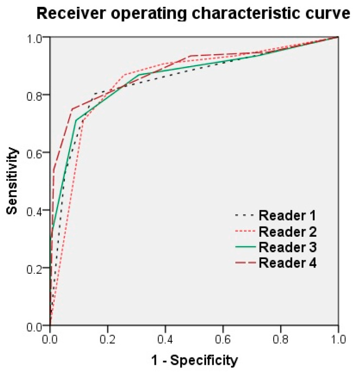
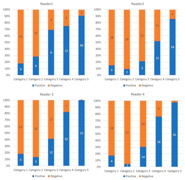
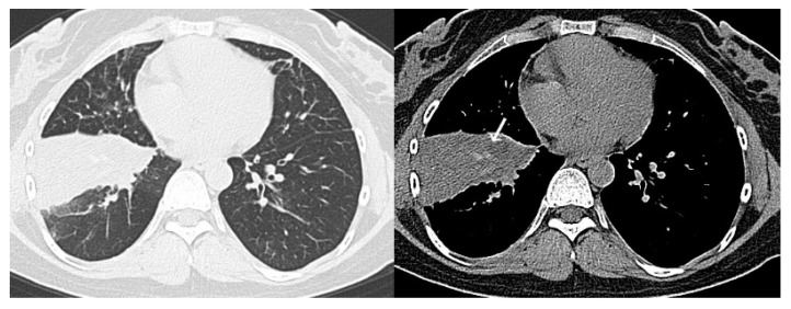
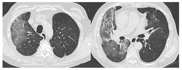
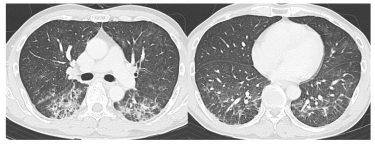
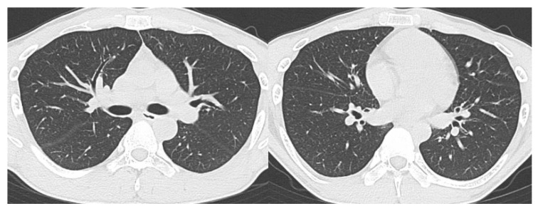
The purpose of this study was to use the Coronavirus Disease 2019 (COVID-19) Reporting and Data System (CO-RADS) to evaluate the chest computed tomography (CT) images of patients suspected of having COVID-19, and to investigate its diagnostic performance and interobserver agreement. The Dutch Radiological Society developed CO-RADS as a diagnostic indicator for assessing suspicion of lung involvement of COVID-19 on a scale of 1 (very low) to 5 (very high). We investigated retrospectively 154 adult patients with clinically suspected COVID-19, between April and June 2020, who underwent chest CT and reverse transcription-polymerase chain reaction (RT-PCR). The patients' average age was 61.3 years (range, 21-93), 101 were male, and 76 were RT-PCR positive. Using CO-RADS, four radiologists evaluated the chest CT images. Sensitivity, specificity, and area under the receiver operating characteristic curve (AUC) were calculated. Interobserver agreement was calculated using the intraclass correlation coefficient (ICC) by comparing the individual reader's score to the median of the remaining three radiologists. The average sensitivity was 87.8% (range, 80.2-93.4%), specificity was 66.4% (range, 51.3-84.5%), and AUC was 0.859 (range, 0.847-0.881); there was no significant difference between the readers (p > 0.200). In 325 (52.8%) of 616 observations, there was absolute agreement among observers. The average ICC of readers was 0.840 (range, 0.800-0.874; p < 0.001). CO-RADS is a categorical taxonomic evaluation scheme for COVID-19 pneumonia, using chest CT images, that provides outstanding performance and from substantial to almost perfect interobserver agreement for predicting COVID-19.

Keywords: CO-RADS; COVID-19; chest imaging; computed tomography; pneumonia; radiology.


Conflict of interest statement

The authors declare no conflict of interest.

Figures

Figure 1
Figure 1
Receiver operating characteristic curve of the four readers. Reader 1, Reader 2, Reader 3, and Reader 4 had an area under the ROC curve (AUC) of 0.847, 0.849, 0.859, and 0.881, respectively. Although the AUC tended to increase with years of radiology experience, there was no significant difference between the readers (p > 0.200).
Figure 2
Figure 2
Percentage and number of COVID19 patients per CO-RADS category. The histogram shows the proportion of COVID19 patients per CO-RADS category. The higher the CO-RADS category, the higher the proportion of COVID-19 positive patients.
Figure 3
Figure 3
Representative computed tomography (CT) images of COVID-19 pneumonia (Case 1). Fifty-two-year-old man. The CT images show multifocal bilateral, peripheral/subpleural ground-glass opacities with consolidations. RT-PCR was positive for SARS-CoV-2. All readers diagnosed it as Category 5. COVID-19, coronavirus disease 2019; CT, computed tomography; RT-PCR, reverse transcription-polymerase chain reaction; SARS-CoV-2, severe acute respiratory syndrome coronavirus 2.
Figure 4
Figure 4
Representative CT images of COVID-19 pneumonia (Case 2). Seventy-seven-year-old female. The CT images show multifocal bilateral, peripheral/subpleural ground-glass opacities with curvilinear bands, and subpleural sparing. RT-PCR was positive for SARS-CoV-2. All readers diagnosed it as Category 5. COVID-19, coronavirus disease 2019; CT, computed tomography; RT-PCR, reverse transcription-polymerase chain reaction; SARS-CoV-2, severe acute respiratory syndrome coronavirus 2.
Figure 5
Figure 5
Representative CT images of a COVID-19-negative patient. Thirty-four-year-old female. The CT images show segmental consolidation with mucus plug (arrow). RT-PCR was negative for SARS-CoV-2 and she was diagnosed with community-acquired pneumonia. All readers diagnosed it as Category 2. COVID-19, coronavirus disease 2019; CT, computed tomography; RT-PCR, reverse transcription-polymerase chain reaction; SARS-CoV-2, severe acute respiratory syndrome coronavirus 2.
Figure 6
Figure 6
Example of a false-positive case (Case 1). Seventy-four-year-old male. The CT images show bilateral ground-glass opacities distributed peripherally to centrally with a right lung predominance. RT-PCR was negative for SARS-CoV-2. She had a history of eosinophilic pneumonia and was diagnosed with relapse of eosinophilic pneumonia. Readers 1, 2, 3, and 4 diagnosed it as Categories 3, 4, 4, and 3, respectively. CT, computed tomography; RT-PCR, reverse transcription-polymerase chain reaction; SARS-CoV-2, severe acute respiratory syndrome coronavirus 2.
Figure 7
Figure 7
Example of a false-positive case (Case 2). Fifty-four-year-old female. The CT images show bilateral, peripheral posterior ground-glass opacities with consolidation with a lower lobe predominance. RT-PCR was negative for SARS-CoV-2. She had a history of medication use and was diagnosed with drug-induced pneumonia. Readers 1, 2, 3, and 4 diagnosed it as Categories 5, 4, 4, and 3, respectively. CT, computed tomography; RT-PCR, reverse transcription-polymerase chain reaction; SARS-CoV-2, severe acute respiratory syndrome coronavirus 2.
Figure 8
Figure 8
Example of a false-negative case. Twenty-five-year-old male. Although RT-PCR was positive for SARS-CoV-2, the CT images show no abnormal findings. All readers diagnosed it as Category 1. CT, computed tomography; RT-PCR, reverse transcription-polymerase chain reaction; SARS-CoV-2, severe acute respiratory syndrome coronavirus 2.

References

    1. Zhu N., Zhang D., Wang W., Li X., Yang B., Song J., Zhao X., Huang B., Shi W., Lu R., et al. A novel coronavirus from patients with pneumonia in China, 2019. N. Engl. J. Med. 2020;382:727–733. doi: 10.1056/NEJMoa2001017. - DOI - PMC - PubMed
    1. Novel Coronavirus (2019-nCoV) Situation Reports. [(accessed on 10 August 2020)]; Available online: https://www.who.int/emergencies/diseases/novel-coronavirus-2019/situatio....
    1. Riccò M., Ferraro P., Gualerzi G., Ranzieri S., Henry B.M., Said Y.B., Pyatigorskaya N.V., Nevolina E., Wu J., Bragazzi N.L., et al. Point-of-care diagnostic tests for detecting SARS-CoV-2 antibodies: A systematic review and meta-analysis of real-world data. J. Clin. Med. 2020;9:1515. doi: 10.3390/jcm9051515. - DOI - PMC - PubMed
    1. Fang Y., Zhang H., Xie J., Lin M., Ying L., Pang P., Ji W. Sensitivity of chest CT for COVID-19: Comparison to RT-PCR. Radiology. 2020:200432. doi: 10.1148/radiol.2020200432. - DOI - PMC - PubMed
    1. Ai T., Yang Z., Hou H., Zhan C., Chen C., Lv W., Tao Q., Sun Z., Xia L. Correlation of chest CT and RT-PCR testing in coronavirus disease 2019 (COVID-19) in China: A report of 1014 cases. Radiology. 2020:200642. doi: 10.1148/radiol.2020200642. - DOI - PMC - PubMed

LinkOut - more resources