A Translational Study on Acute Traumatic Brain Injury: High Incidence of Epileptiform Activity on Human and Rat Electrocorticograms and Histological Correlates in Rats
- PMID: 32825101
- PMCID: PMC7565553
- DOI: 10.3390/brainsci10090570
A Translational Study on Acute Traumatic Brain Injury: High Incidence of Epileptiform Activity on Human and Rat Electrocorticograms and Histological Correlates in Rats
Abstract
Background: In humans, early pathological activity on invasive electrocorticograms (ECoGs) and its putative association with pathomorphology in the early period of traumatic brain injury (TBI) remains obscure.
Methods: We assessed pathological activity on scalp electroencephalograms (EEGs) and ECoGs in patients with acute TBI, early electrophysiological changes after lateral fluid percussion brain injury (FPI), and electrophysiological correlates of hippocampal damage (microgliosis and neuronal loss), a week after TBI in rats.
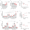
Results: Epileptiform activity on ECoGs was evident in 86% of patients during the acute period of TBI, ECoGs being more sensitive to epileptiform and periodic discharges. A "brush-like" ECoG pattern superimposed over rhythmic delta activity and periodic discharge was described for the first time in acute TBI. In rats, FPI increased high-amplitude spike incidence in the neocortex and, most expressed, in the ipsilateral hippocampus, induced hippocampal microgliosis and neuronal loss, ipsilateral dentate gyrus being most vulnerable, a week after TBI. Epileptiform spike incidence correlated with microglial cell density and neuronal loss in the ipsilateral hippocampus.
Conclusion: Epileptiform activity is frequent in the acute period of TBI period and is associated with distant hippocampal damage on a microscopic level. This damage is probably involved in late consequences of TBI. The FPI model is suitable for exploring pathogenetic mechanisms of post-traumatic disorders.
Keywords: electrocorticograms; epileptiform discharges; hippocampus; local field potentials; microglia; neocortex; neurodegeneration; post-traumatic epilepsy; traumatic brain injury.
Conflict of interest statement
The authors declare no conflict of interest.
Figures


Similar articles
-
[Early electrophysiological consequences of dosed traumatic-brain injury in rats].Zh Nevrol Psikhiatr Im S S Korsakova. 2018;118(10. Vyp. 2):21-26. doi: 10.17116/jnevro201811810221. Zh Nevrol Psikhiatr Im S S Korsakova. 2018. PMID: 30698540 Russian.
-
Differential early effects of traumatic brain injury on spike-wave discharges in Sprague-Dawley rats.Neurosci Res. 2021 May;166:42-54. doi: 10.1016/j.neures.2020.05.005. Epub 2020 May 24. Neurosci Res. 2021. PMID: 32461140
-
Neuroinflammatory Cytokine Response, Neuronal Death, and Microglial Proliferation in the Hippocampus of Rats During the Early Period After Lateral Fluid Percussion-Induced Traumatic Injury of the Neocortex.Mol Neurobiol. 2022 Feb;59(2):1151-1167. doi: 10.1007/s12035-021-02668-4. Epub 2021 Dec 2. Mol Neurobiol. 2022. PMID: 34855115
-
Acute Non-Convulsive Status Epilepticus after Experimental Traumatic Brain Injury in Rats.J Neurotrauma. 2019 Jun;36(11):1890-1907. doi: 10.1089/neu.2018.6107. Epub 2019 Feb 25. J Neurotrauma. 2019. PMID: 30543155 Free PMC article.
-
The Impact of Previous Physical Training on Redox Signaling after Traumatic Brain Injury in Rats: A Behavioral and Neurochemical Approach.J Neurotrauma. 2016 Jul 15;33(14):1317-30. doi: 10.1089/neu.2015.4068. Epub 2016 Apr 15. J Neurotrauma. 2016. PMID: 26651029
Cited by
-
Robust, long-term video EEG monitoring in a porcine model of post-traumatic epilepsy.eNeuro. 2022 Jun 10;9(4):ENEURO.0025-22.2022. doi: 10.1523/ENEURO.0025-22.2022. Online ahead of print. eNeuro. 2022. PMID: 35697513 Free PMC article.
-
Neuroinflammation and Neuronal Loss in the Hippocampus Are Associated with Immediate Posttraumatic Seizures and Corticosterone Elevation in Rats.Int J Mol Sci. 2021 May 30;22(11):5883. doi: 10.3390/ijms22115883. Int J Mol Sci. 2021. PMID: 34070933 Free PMC article.
-
Spike-induced cytoarchitectonic changes in epileptic human cortex are reduced via MAP2K inhibition.Brain Commun. 2024 Apr 29;6(3):fcae152. doi: 10.1093/braincomms/fcae152. eCollection 2024. Brain Commun. 2024. PMID: 38741662 Free PMC article.
-
Resilience of Spontaneously Hypertensive Rats to Secondary Insults After Traumatic Brain Injury: Immediate Seizures, Survival, and Stress Response.Int J Mol Sci. 2025 Jan 19;26(2):829. doi: 10.3390/ijms26020829. Int J Mol Sci. 2025. PMID: 39859543 Free PMC article.
References
-
- Reilly P. Progress in Brain Research. Volume 161. Elsevier; Amsterdam, The Netherlands: 2007. The impact of neurotrauma on society: An international perspective; pp. 3–9. - PubMed
-
- Pitkänen A., Kyyriäinen J., Andrade P., Pasanen L., Ndode-Ekane X.E. Models of Seizures and Epilepsy. Elsevier; Amsterdam, The Netherlands: 2017. Epilepsy After Traumatic Brain Injury; pp. 661–681.
Grants and funding
LinkOut - more resources
Full Text Sources

