Review
. 2020 Aug 19;9(9):1925.
doi: 10.3390/cells9091925.

The Diversity of Muscles and Their Regenerative Potential across Animals

Affiliations
Review

The Diversity of Muscles and Their Regenerative Potential across Animals

Letizia Zullo et al. Cells. .

Abstract

Cells with contractile functions are present in almost all metazoans, and so are the related processes of muscle homeostasis and regeneration. Regeneration itself is a complex process unevenly spread across metazoans that ranges from full-body regeneration to partial reconstruction of damaged organs or body tissues, including muscles. The cellular and molecular mechanisms involved in regenerative processes can be homologous, co-opted, and/or evolved independently. By comparing the mechanisms of muscle homeostasis and regeneration throughout the diversity of animal body-plans and life cycles, it is possible to identify conserved and divergent cellular and molecular mechanisms underlying muscle plasticity. In this review we aim at providing an overview of muscle regeneration studies in metazoans, highlighting the major regenerative strategies and molecular pathways involved. By gathering these findings, we wish to advocate a comparative and evolutionary approach to prompt a wider use of "non-canonical" animal models for molecular and even pharmacological studies in the field of muscle regeneration.

Keywords: differentiation; evolution; metazoans; muscle precursors; myogenesis; regenerative medicine; transdifferentiation.


Conflict of interest statement

The authors declare no conflict of interest.

Figures

Figure 1
Figure 1
Tree of the animals’ clades treated in this review (in brackets the corresponding section numbers).
Figure 2
Figure 2
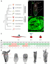
The epitheliomuscular system and regenerative capacity of the anthozoan cnidarian Nematostella vectensis. (A) Schematic representation of the relationship between the main cnidarian lineages and the phylogenetic position of Nematostella vectensis (Anthozoa, Hexacorallia). (B) The upper panel shows the muscle network of nematostella in a fixed MyHC1::mCherry transgene [30] labeling the retractor muscles, co-stained with phalloidin thus showing the entire muscle network in green. (ten) tentacles, (*) mouth, (pha) pharynx, (bc) body column, (ph) physa, (m) mesenteries, (rm) retractor muscles, (pm) parietal muscles. (B’) Magnification of a body column region to highlight the orientation of the muscle fibers. (tmf) transverse muscle fibers, (lmf) longitudinal muscle fibers. (C) Three epitheliomuscular cell types have been identified in nematostella; they vary in their apical and basal cell junctions as well as their localizations within the body [31]. (D) Overview of the known bilateral myogenic factors identified in nematostella. (Present) indicates that the gene has been identified in the genome, (Role) indicates a myogenic role (or not) of this gene in nematostella; (pot. yes), indicates evidence of a myogenic role based on functional experiments or gene expression. (pot. not), indicates evidence of a non-myogenic role based on functional experiments or gene expression. (n/a) data not available. References cited: (a) [32], (b) [33], (c) [34], (d) [35], (e) [36], (f) [37], (g) [38], (h) [39], (i) [40]. (E) Oral regeneration of lost body parts after sub-pharyngeal amputation (red dashed line) is completed after 120 h post amputation and reforms a fully functional organism. Animals were fixed at various time points during regeneration and stained with phalloidin to show f-actin filaments (black). Elements of the figure are extracted from [28,41].
Figure 3
Figure 3
Timing and principal steps of regeneration in B. lanceolatum and B. japonicum. (A) Enlargement of the most anterior end of a Branchiostoma lanceolatum adult showing the oral cirri (oc). (B) Post-anal tail of the same animal. my, myomeres; nc, nerve cord; n, notochord. (C) B. lanceolatum individual collected in Banyuls-sur-Mer, France. (D) Schematic overview of the series of events occurring during tail regeneration in the B. lanceolatum and B. japonicum. dpa, days post-amputation.
Figure 4
Figure 4
Ciona embryogenesis and regeneration. (A) Simplified scheme of muscle development during ciona embryogenesis and metamorphoses. (modified from [163]). (B) Scheme of oral siphon regeneration in ciona (modified from [164]). (C) Simplified scheme of myogenesis during budding in Botryllus schlosseri. FHF: first heart field; SHF: second heart field; OS: oral siphon; AS: atrial siphon; LoM: longitudinal muscles; SC: stem cells.
Figure 5
Figure 5
Regeneration in zebrafish. Schematic drawing of a zebrafish adult showing organs used for regeneration studies.

References

    1. Ciciliot S., Schiaffino S. Regeneration of mammalian skeletal muscle. Basic mechanisms and clinical implications. Curr. Pharm. Des. 2010;16:906–914. doi: 10.2174/138161210790883453. - DOI - PubMed
    1. Musaro A. The basis of muscle regeneration. Adv. Biol. 2014;2014:1–16. doi: 10.1155/2014/612471. - DOI
    1. Maden M. The evolution of regeneration—Where does that leave mammals? Int. J. Dev. Biol. 2018;62:369–372. doi: 10.1387/ijdb.180031mm. - DOI - PubMed
    1. Tiozzo S., Copley R. Reconsidering regeneration in metazoans: An evo-devo approach. Front. Ecol. Evol. 2015;3:67. doi: 10.3389/fevo.2015.00067. - DOI
    1. Steinmetz P.R., Kraus J.E., Larroux C., Hammel J.U., Amon-Hassenzahl A., Houliston E., Worheide G., Nickel M., Degnan B.M., Technau U. Independent evolution of striated muscles in cnidarians and bilaterians. Nature. 2012;487:231–234. doi: 10.1038/nature11180. - DOI - PMC - PubMed

Publication types

LinkOut - more resources