Genetic and Phenotypic Characterization of a Rabies Virus Strain Isolated from a Dog in Tokyo, Japan in the 1940s
- PMID: 32825306
- PMCID: PMC7552007
- DOI: 10.3390/v12090914
Genetic and Phenotypic Characterization of a Rabies Virus Strain Isolated from a Dog in Tokyo, Japan in the 1940s
Abstract
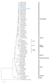
The rabies virus strain Komatsugawa (Koma), which was isolated from a dog in Tokyo in the 1940s before eradication of rabies in Japan in 1957, is known as the only existent Japanese field strain (street strain). Although this strain potentially provides a useful model to study rabies pathogenesis, little is known about its genetic and phenotypic properties. Notably, this strain underwent serial passages in rodents after isolation, indicating the possibility that it may have lost biological characteristics as a street strain. In this study, to evaluate the utility of the Koma strain for studying rabies pathogenesis, we examined the genetic properties and in vitro and in vivo phenotypes. Genome-wide genetic analyses showed that, consistent with previous findings from partial sequence analyses, the Koma strain is closely related to a Russian street strain within the Arctic-related phylogenetic clade. Phenotypic examinations in vitro revealed that the Koma strain and the representative street strains are less neurotropic than the laboratory strains. Examination by using a mouse model demonstrated that the Koma strain and the street strains are more neuroinvasive than the laboratory strains. These findings indicate that the Koma strain retains phenotypes similar to those of street strains, and is therefore useful for studying rabies pathogenesis.
Keywords: Arctic-related clade; Komatsugawa; fixed virus; pathogenesis; rabies virus; street virus.
Conflict of interest statement
The authors declare no conflict of interest.
Figures


Similar articles
-
Establishment of a reverse genetics system for rabies virus strain Komatsugawa.J Vet Med Sci. 2022 Nov 7;84(11):1508-1513. doi: 10.1292/jvms.22-0254. Epub 2022 Sep 29. J Vet Med Sci. 2022. PMID: 36171109 Free PMC article.
-
[Phylogenetic analysis of rabies viruses isolated from animals in Tokyo in the 1950s].Kansenshogaku Zasshi. 2011 May;85(3):238-43. doi: 10.11150/kansenshogakuzasshi.85.238. Kansenshogaku Zasshi. 2011. PMID: 21706842 Japanese.
-
Molecular characterization of a rabies virus isolate from a rabid dog in Hanzhong District, Shaanxi Province, China.Arch Virol. 2014 Jun;159(6):1481-6. doi: 10.1007/s00705-013-1941-y. Epub 2013 Dec 19. Arch Virol. 2014. PMID: 24352434
-
Genetic characterization of rabies virus isolates in Korea.Virus Genes. 2005 May;30(3):341-7. doi: 10.1007/s11262-005-6777-4. Virus Genes. 2005. PMID: 15830152
-
[Sequencing and analysis of complete genome of rabies viruses isolated from Chinese Ferret-Badger and dog in Zhejiang province].Bing Du Xue Bao. 2010 Jan;26(1):45-52. Bing Du Xue Bao. 2010. PMID: 20329558 Chinese.
Cited by
-
Point Mutations in the Glycoprotein Ectodomain of Field Rabies Viruses Mediate Cell Culture Adaptation through Improved Virus Release in a Host Cell Dependent and Independent Manner.Viruses. 2021 Oct 3;13(10):1989. doi: 10.3390/v13101989. Viruses. 2021. PMID: 34696419 Free PMC article.
-
Reverse genetic approaches allowing the characterization of the rabies virus street strain belonging to the SEA4 subclade.Sci Rep. 2024 Aug 9;14(1):18509. doi: 10.1038/s41598-024-69613-y. Sci Rep. 2024. PMID: 39122768 Free PMC article.
-
The Amino Acid at Position 95 in the Matrix Protein of Rabies Virus Is Involved in Antiviral Stress Granule Formation in Infected Cells.J Virol. 2022 Sep 28;96(18):e0081022. doi: 10.1128/jvi.00810-22. Epub 2022 Sep 7. J Virol. 2022. PMID: 36069552 Free PMC article.
-
Establishment of a reverse genetics system for rabies virus strain Komatsugawa.J Vet Med Sci. 2022 Nov 7;84(11):1508-1513. doi: 10.1292/jvms.22-0254. Epub 2022 Sep 29. J Vet Med Sci. 2022. PMID: 36171109 Free PMC article.
-
AP3B1 facilitates PDIA3/ERP57 function to regulate rabies virus glycoprotein selective degradation and viral entry.Autophagy. 2024 Dec;20(12):2785-2803. doi: 10.1080/15548627.2024.2390814. Epub 2024 Aug 17. Autophagy. 2024. PMID: 39128851 Free PMC article.
References
-
- Jackson A.C., Fu Z.F. Rabies. Elsevier Inc.; Amsterdam, The Netherlands: 2013. Pathogenesis; pp. 299–349.
-
- WHO/Department of Control of Neglected Tropical Diseases Human Rabies Transmitted by Dogs: Current Status of Global Data, 2015. Wkly. Epidemiol. Rec. 2016;2:13–20. - PubMed
-
- WHO/Department of Control of Neglected Tropical Diseases Rabies Vaccines: WHO Position Paper–April 2018. Wkly. Epidemiol. Rec. 2018;16:201–220.
-
- Jackson A.C., Fu Z.F. Rabies. Elsevier Inc.; Amsterdam, The Netherlands: 2013. Rabies virus; pp. 17–60.
Publication types
MeSH terms
Substances
LinkOut - more resources
Full Text Sources
Medical

