. 2020 Aug 20;8(9):1263.
doi: 10.3390/microorganisms8091263.

Atypical Membrane-Anchored Cytokine MIF in a Marine Dinoflagellate

Affiliations

Atypical Membrane-Anchored Cytokine MIF in a Marine Dinoflagellate

Maëlle Jaouannet et al. Microorganisms. .

Abstract

Macrophage Migration Inhibitory Factors (MIF) are pivotal cytokines/chemokines for vertebrate immune systems. MIFs are typically soluble single-domain proteins that are conserved across plant, fungal, protist, and metazoan kingdoms, but their functions have not been determined in most phylogenetic groups. Here, we describe an atypical multidomain MIF protein. The marine dinoflagellate Lingulodinium polyedra produces a transmembrane protein with an extra-cytoplasmic MIF domain, which localizes to cell-wall-associated membranes and vesicular bodies. This protein is also present in the membranes of extracellular vesicles accumulating at the secretory pores of the cells. Upon exposure to biotic stress, L. polyedra exhibits reduced expression of the MIF gene and reduced abundance of the surface-associated protein. The presence of LpMIF in the membranes of secreted extracellular vesicles evokes the fascinating possibility that LpMIF may participate in intercellular communication and/or interactions between free-living organisms in multispecies planktonic communities.

Keywords: Lingulodinium polyedra; MIF; dinoflagellate; secretion; stress response; transmembrane protein.


Conflict of interest statement

The authors declare no conflict of interest.

Figures

Figure 1
Figure 1
Lingulodinium polyedra expresses transmembrane Migration Inhibitory Factors (MIF). (a) Alignment of the L. polyedra MIF (LpMIF) sequence with human MIF (dark green). Amino acid numbers of the human MIF are given below the sequence. Hyphens (-) indicate a gap in the respective sequence. Amino acids shown to determine the tautomerase activity (■) and oxidoreductase activity (CXXC motif; O) in human MIF are shown by symbols above the sequence. The peptide identified by mass spectrometry is indicated by a line above the LpMIF sequence. (b) Location (LpMIF amino-acid number) and orientation of the transmembrane helices predicted by five prediction tools, where « i » refers to inside (cytoplasmic) and « o » to « outside » (non-cytoplasmic). (c) Schematic representation of the predicted LpMIF topology. The transmembrane domains are represented in blue and the MIF domain in green. The membrane is represented as a lipidic bilayer. (d) Western blot analysis of the membrane fraction (Mf) of L. polyedra proteins. The membrane extract (10 µg of total proteins) reveals a major protein with an apparent molecular mass of about 60 kDa. The presence of LpMIF in the most prominent band of the membrane fraction was confirmed by mass spectrometry (see 1a). (e,f) Subcellular localization of LpMIF fused to GFP in the N-terminal region, with (e) or without (f) the transmembrane domains in Nicotiana benthamiana. Leaves were inoculated with A. tumefaciens harboring the LpMIF-GFP constructs, 2 days prior to observations. Complete LpMIF proteins localize to the membranes of vesicles and the plasma membrane (PM) (e), while LpMIF without the transmembrane domains (in f) shows a nucleo-cytoplasmic expression in N. benthamiana cells, which is typical for small, soluble proteins. (N) nuclei, (Cyt) cytoplasm.
Figure 2
Figure 2
Subcellular localization of LpMIF. (a,b) TEM of immunogold-labeled L. polyedra cells showing the accumulation of gold particles (black dots) in membranes of the vesicular bodies (VB) and in the theca. Note that one of the vesicular bodies (indicated by a star) appears to be in a process of fusion with the cell wall. (c,d) Immunolocalization of LpMIF by confocal microscopy. LpMIF is labelled in green in (c,d), while the nucleus (N) appears in blue in (d) (DAPI staining). The overall structure of L. polyedra is visible under UV light due to autofluorescence in (d), and shows the shape of the thecal plates, the apical pore (AP), and the thecal pores (TP). Note the accumulation of LpMIF at the apical pore (AP) and the thecal pores (TP). (e,f) Scanning electron micrographs of L. polyedra showing the AP and TP. An enlargement of a TP in (f) shows the inner pore (P). (g,h) Representative pictures of L. polyedra cells that have been immunofluorescence-labelled under mild washing and fixing conditions, revealing the accumulation of MIF-labelled extracellular vesicles. Here, an apical view of the cell shows labelled vesicles surrounding the cell (g) and a lateral view illustrate the commonly observed accumulation of labelled mucus at the apical pore (AP). Control samples exposed to pre-immune serum or no primary antibody did not show any fluorescent signal (Figure S1e,f). Please see Supplementary Materials for procedure description.
Figure 3
Figure 3
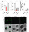
LpMIF expression during a stress response. (a) The exposure of L. polyedra to copepods decreases photosynthetic activity as a stress response. Chlorophyll fluorescence parameters were measured with a Multi-Color Pulse-Amplitude-Modulated fluorometer (Heinz Walz Gmbh, Effeltrich, Germany). Photosystem II activity was calculated from the ratio of variable fluorescence to maximum chlorophyll fluorescence (Fv/Fm). Values are means (+/− SEM) from three independent measurements/experiments. Asterisks indicate statistical differences (*** p < 0.001), according to Kruskal-Wallis test. (b) Relative expression ratios (normalized to control) of LpMIF in L. polyedra cells before (control), during (stress), and after (post-stress) exposure to copepods. Asterisks indicate statistical differences (*** p < 0.001), according to Kruskal-Wallis test. (c) Relative fluorescence of MIF-labelled L. polyedra cells expressed as the fluorescent area/total area, of cells before (control), during (stress), and after (post-stress) exposure to copepods (n = 9 per sample; Kruskal-Wallis test: ** = p ≤ 0.01, *** = p ≤ 0.001). (d) Representative pictures of MIF-labelled L. polyedra before (control), during (stress), and after (post-stress) exposure to copepods. Images in the upper row are fluorescence micrographs (Anti-MIF) that were merged in the lower row with transmission light micrographs (Merged). Negative control samples directly exposed to the secondary antibody are shown in the Figure S2.

References

    1. Lue H., Kleemann R., Calandra T., Roger T., Bernhagen J. Macrophage migration inhibitory factor (MIF): Mechanisms of action and role in disease. Microbes Infect. 2002;4:449–460. doi: 10.1016/S1286-4579(02)01560-5. - DOI - PubMed
    1. Günther S., Fagone P., Jalce G., Atanasov A.G., Guignabert C., Nicoletti F. Role of MIF and D-DT in immune-inflammatory, autoimmune, and chronic respiratory diseases: From pathogenic factors to therapeutic targets. Drug Discov. Today. 2019;24:428–439. doi: 10.1016/j.drudis.2018.11.003. - DOI - PubMed
    1. Harris J., VanPatten S., Deen N.S., Al-Abed Y., Morand E. Rediscovering MIF: New tricks for an old cytokine. Trends Immunol. 2019;40:447–462. doi: 10.1016/j.it.2019.03.002. - DOI - PubMed
    1. Morand E.F., Leech M., Bernhagen J. MIF: A new cytokine link between rheumatoid arthritis and atherosclerosis. Nat. Rev. Drug Discov. 2006;5:399–411. doi: 10.1038/nrd2029. - DOI - PubMed
    1. Bernhagen J., Calandra T., Bucala R. Regulation of the immune response by macrophage migration inhibitory factor: Biological and structural features. J. Mol. Med. 1998;76:151–161. doi: 10.1007/s001090050204. - DOI - PubMed

LinkOut - more resources