Review
. 2020 Aug 20;21(17):5994.
doi: 10.3390/ijms21175994.

Phage Display to Augment Biomaterial Function

Affiliations
Review

Phage Display to Augment Biomaterial Function

Thomas A Davidson et al. Int J Mol Sci. .

Abstract

Biomaterial design relies on controlling interactions between materials and their biological environments to modulate the functions of proteins, cells, and tissues. Phage display is a powerful tool that can be used to discover peptide sequences with high affinity for a desired target. When incorporated into biomaterial design, peptides identified via phage display can functionalize material surfaces to control the interaction between a biomaterial and its local microenvironment. A targeting peptide has high specificity for a given target, allowing for homing a specific protein, cell, tissue, or other material to a biomaterial. A functional peptide has an affinity for a given protein, cell, or tissue, but also modulates its target's activity upon binding. Biomaterials can be further enhanced using a combination of targeting and/or functional peptides to create dual-functional peptides for bridging two targets or modulating the behavior of a specific protein or cell. This review will examine current and future applications of phage display for the augmentation of biomaterials.

Keywords: biomaterials; dual-functioning peptides; peptides; phage display; tissue engineering.


Conflict of interest statement

The authors declare no conflict of interest.

Figures
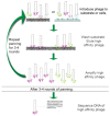
Figure 1
Figure 1
Schematic of panning stage of phage display. Phages are introduced to either a substrate or specific cell population followed by washing away non-adherent phages and eluting high affinity phages. High-affinity phages are amplified and the process is repeated at least 3 times to reach a consensus sequence. Image used with permission from Springer.
Figure 2
Figure 2
Wide-field endoscopy images acquired after in vivo incubation (5 min) of fluorescently labeled peptide QPIHPNNM in a genetic mouse model of adenoma formation in the colon. The left column shows white light images and the right column shows fluorescence images for binding of (A) QPIHPNNM to colon tissue with multiple adenomas, (B) QPIHPNNM to colon tissue with one adenoma, (C) control peptide GGGAGGGA to colon tissue with multiple adenomas, and (D) QPIHPNNM to lumen of colon tissue of a wild-type (WT) mouse. In the model shown, CPC:Apc, Cre recombinase is linked to the Cdx2 promoter and floxed APC (adenomatous polyposis coli). Images modified from [10] and used with permission from PLoS ONE.
Figure 3
Figure 3
Representation of steps in the phage display process to narrow the phage library down to only peptides with high affinity to the target substrate or cell population. Image modified from [7] and used with permission from Springer.
Figure 4
Figure 4
These graphs demonstrate how (A) cost and (B) time necessary for phage sequencing decreases when using next-generation sequencing (blue) over traditional Sanger sequencing (red) for increasing numbers of sequences. Images modified from [15] and used with permission from PLoS ONE.
Figure 5
Figure 5
Representative immunofluorescence images of fluorescently labeled peptide CARG (FAM-CARG, green) homing and binding to lungs infected with Staphylococcus aureus (red; top row) and sham-infected lungs (bottom row) with DAPI (blue) staining of nuclei. In each row, the leftmost image shows lung tissue labelled with FAM-CARG (green) and DAPI (blue); the white box outlines the region of interest (ROI) examined at higher magnification (scale bar = 50um). In each row, the ROI is shown with individual fluorescent channels FAM-CARG (middle left) and S. aureus (middle right), and all fluorescent channels (right). FAM-CARG was intravenously injected into tails of infected mice and allowed to circulate for 30 min prior to sacrifice and histological examination (n = 3–5 mice per group with 5 histological samples examined per lung). Images modified from [43] and used with permission from Nature Biomedical Engineering.
Figure 6
Figure 6
Dual-functioning peptide schematic with a targeting peptide (blue) attached to a substrate (dark grey) connected to a functional or specific cell targeting peptide (yellow) via spacer amino acids (light grey). Image modified from [44] and used with permission from Advanced Materials.
Figure 7
Figure 7
(A) Half-cell detachment force (τ50) measured from peptide-coated apatite films of human bone marrow stem cells, murine bone marrow stem cells (mBMSCs), induced pluripotent stem cell (iPS)-derived mesenchymal stem cells (MSCs), pre-osteoblastic MCTC3s, and murine dermal fibroblasts (MDFs). (B) Confocal microscopy images taken at 40x show increased spreading of mBMSCs and iPS-MSCs on DPI-VTK-coated apatite films and increased spreading of MCTC3 and MDFs on RGD-VTK-coated apatite films. (C) Quantitative analysis of cell spreading via ImageJ on peptide-coated apatite films. (D) Total number of cells over time, reaching saturation at day 10. * denotes significance compared to apatite control and tissue culture polystyrene (TCPS). * denotes significance compared to apatite control. ** denotes significance compared to VTK. (EH) Relative gene expression of osteogenic markers (Runx2, OSX, ALP, and OCN) normalized to day 0 and GAPDH of iPS-MSCs on biomimetic apatite and peptide coated apatite. Images used with permission from Taylor & Francis.

References

    1. Smith G.P. Filamentous fusion phage: Novel expression vectors that display cloned antigens on the virion surface. Science. 1985;228:1315–1317. doi: 10.1126/science.4001944. - DOI - PubMed
    1. Sidhu S.S. Current Opinion in Biotechnology. Elsevier Ltd.; Amsterdam, The Netherlands: 2000. Phage display in pharmaceutical biotechnology; pp. 610–616. - DOI - PubMed
    1. Kehoe J.W., Kay B.K. Filamentous phage display in the new millennium. Chem. Rev. 2005;105:4056–4072. doi: 10.1021/cr000261r. - DOI - PubMed
    1. Newton J.R., Deutscher S.L. In Vivo bacteriophage display for the discovery of novel peptide-based tumor-targeting agents. Methods Mol. Biol. 2009;504:275–290. doi: 10.1007/978-1-60327-569-9_17. - DOI - PubMed
    1. Rosenberg A., Griffin K., Studier E.W., Mccormick M., Berg J., Novy R., Mierendorf R. T7 Select phage display system: A powerful new protein display system based on bacteriophage T7. Innovations. 1996;6:1–6.

LinkOut - more resources