. 2020 Aug 21;12(9):538.
doi: 10.3390/toxins12090538.

Primary Sequence and 3D Structure Prediction of the Plant Toxin Stenodactylin

Affiliations

Primary Sequence and 3D Structure Prediction of the Plant Toxin Stenodactylin

Rosario Iglesias et al. Toxins (Basel). .

Abstract

Stenodactylin is one of the most potent type 2 ribosome-inactivating proteins (RIPs); its high toxicity has been demonstrated in several models both in vitro and in vivo. Due to its peculiarities, stenodactylin could have several medical and biotechnological applications in neuroscience and cancer treatment. In this work, we report the complete amino acid sequence of stenodactylin and 3D structure prediction. The comparison between the primary sequence of stenodactylin and other RIPs allowed us to identify homologies/differences and the amino acids involved in RIP toxic activity. Stenodactylin RNA was isolated from plant caudex, reverse transcribed through PCR and the cDNA was amplificated and cloned into a plasmid vector and further analyzed by sequencing. Nucleotide sequence analysis showed that stenodactylin A and B chains contain 251 and 258 amino acids, respectively. The key amino acids of the active site described for ricin and most other RIPs are also conserved in the stenodactylin A chain. Stenodactylin amino acid sequence shows a high identity degree with volkensin (81.7% for A chain, 90.3% for B chain), whilst when compared with other type 2 RIPs the identity degree ranges from 27.7 to 33.0% for the A chain and from 42.1 to 47.7% for the B chain.

Keywords: 3D structure; plant toxin; primary sequence; ribosome-inactivating protein; stenodactylin; toxic lectin.


Conflict of interest statement

The authors declare no conflict of interest.

Figures
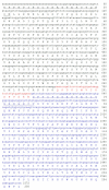
Figure 1
Figure 1
Full length sequence and derived amino acid sequence of the stenodactylin gene. The A chain is presented in black; the B chain is presented in blue and the sequence of the connecting peptides is presented in red. The amino acid sequences obtained by Edman degradation, as described in [7], are underlined. Numbering refers to the position of the amino acids in the mature A and B chains. The cDNA sequence for stenodactylin was submitted to GenBank (accession number: MT580807). The letter “n” means “unknown nucleotide residue”, being the amino acid sequence obtained exclusively by Edman degradation.
Figure 2
Figure 2
Secondary structure analysis of stenodactylin A and B chains. The secondary structure motifs were predicted using the PSIPRED Protein Structure Prediction Server. The predicted helix (H, pink) and strand (E, yellow) structure elements and randomly structured coil regions (C) of the target sequences are displayed according to the symbols shown in the legend. The confidence levels of the prediction are reported in the figure.
Figure 3
Figure 3
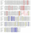
Alignment between stenodactylin and volkensin (GenBank CAD61022). Identical residues (*), conserved substitutions (:) and semiconserved substitutions (.) are reported. The A and B chains are presented in black; the sequence of the linker peptides is presented in gray. The putative amino acids that are present in the active site pocket (boxed in red) or in the galactoside-binding sites (boxed in blue), those involved in substrate binding or catalysis (highlighted in red), those involved in sugar binding (highlighted in blue), and those involved in disulfide bridges (highlighted in yellow) are represented, and they were assigned by comparison with the structure of ricin (accession no. 2AAI, 3RTI and 3RTJ). The dash indicates a gap introduced into the sequences to maximize alignments.
Figure 4
Figure 4
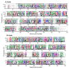
Structure of stenodactylin compared with ricin. (a) Amino acid sequence alignment of the A and B chains of stenodactylin and ricin. The β strands (blue), the α helices (red) and the cysteines involved in the disulfide bonds (highlighted in yellow) are indicated. The helices are labelled A to I and the strands of the β sheets are labelled a to h in the A chain. The domains and subdomains in the B chain are also indicated. Identical residues (*), conserved substitutions (:) and semiconserved substitutions (.) are reported. The cartoons represent the different structural motifs in both A and B chains. (b) Three-dimensional structure of stenodactylin compared with ricin (Protein Data Bank accession no. 2AAI). The three-dimensional structural modelling was carried out on the I-TASSER server and the figure was generated using Discovery Studio 2016. The α helices (red), the β chains (cyan), and the coils (grey) are represented. The helices are labelled A to I and the strands of the β sheets are labelled a to h in the A chain. The structural domains and subdomains in the B chain are also indicated. Arrows indicate the position of the disulfide bond linking A and B chains.
Figure 5
Figure 5
Structure of stenodactylin B chain. The three-dimensional structural modelling was carried out on the I-TASSER server and the figure was generated using Discovery Studio 2016. The structural domains and subdomains in the B chain are indicated.
Figure 6
Figure 6
Protein sequence alignment of stenodactylin with volkensin (accession no. CAD61022), ricin (accession no. P02879), abrin a (accession no. P11140), cinnamomin I (accession no. AAF68978), viscumin (accession no. P81446), riproximin (accession no. CAJ38823), ebulin l (accession no. CAC33178), and nigrin b (accession no. P33183). Identical residues (*), conserved substitutions (:) and semiconserved substitutions (.) are reported. The A and B chains are presented in black and the sequence of the connecting peptide is presented in red. The putative amino acids that are present in the active site pocket (boxed in red) or in the galactoside-binding sites (boxed in blue), those involved in substrate binding or catalysis (highlighted in red), those involved in sugar binding (highlighted in blue), and those involved in disulphide bridges (highlighted in yellow) are represented, and they were assigned by comparison with the structure of ricin (accession no. 2AAI, 3RTI and 3RTJ). Dashes denote gaps introduced into the sequences to maximize alignments.
Figure 7
Figure 7
Sequence logos of the A and B chains of type 2 RIPs. The sequence logo representation of the alignment of the A and B-chain sequences from 46 representative type 2 RIPs belonging to 28 plant species was created as indicated in the “Materials and Methods” section. Letter height is proportional to the frequency of that amino acid at that position in the alignment respect to all the amino acids; letter width is proportional to the frequency of that amino acid but includes gaps. The sequence of stenodactylin is indicated above the logos.
Figure 8
Figure 8
Molecular phylogenetic analysis by the Maximum Likelihood method of representative A and B-chain type 2 RIPs. The evolutionary history was inferred as indicated in the “Materials and Methods” section. The sequences of representative type 1 RIPs and monomeric lectins were used as the outgroup for the A and B chains, respectively. The name of the RIP (if any), the species and the accession number are indicated. All the sequences were retrieved and processed as indicated in the “Materials and Methods” section.

Similar articles

Cited by

References

    1. Bolognesi A., Bortolotti M., Maiello S., Battelli M.G., Polito L. Ribosome-Inactivating Proteins from Plants: A Historical Overview. Molecules. 2016;21:1627. doi: 10.3390/molecules21121627. - DOI - PMC - PubMed
    1. Polito L., Bortolotti M., Maiello S., Battelli M.G., Bolognesi A. Plants Producing Ribosome-Inactivating Proteins in Traditional Medicine. Molecules. 2016;21:1560. doi: 10.3390/molecules21111560. - DOI - PMC - PubMed
    1. Bortolotti M., Mercatelli D., Polito L. Momordica charantia, a Nutraceutical Approach for Inflammatory Related Diseases. Front. Pharmacol. 2019;10:486. doi: 10.3389/fphar.2019.00486. - DOI - PMC - PubMed
    1. Barbieri L., Valbonesi P., Bonora E., Gorini P., Bolognesi A., Stirpe F. Polynucleotide:adenosine glycosidase activity of ribosome-inactivating proteins: Effect on DNA, RNA and poly(A) Nucleic Acids Res. 1997;25:518–522. doi: 10.1093/nar/25.3.518. - DOI - PMC - PubMed
    1. Battelli M.G., Barbieri L., Bolognesi A., Buonamici L., Valbonesi P., Polito L., Van Damme E.J.M., Peumans W.J., Stirpe F. Ribosome-inactivating lectins with polynucleotide:adenosine glycosidase activity. FEBS Lett. 1997;408:355–359. doi: 10.1016/S0014-5793(97)00463-8. - DOI - PubMed

Publication types

LinkOut - more resources