

Effect of niacin monotherapy on high density lipoprotein composition and function
- PMID: 32825822
- PMCID: PMC7441610
- DOI: 10.1186/s12944-020-01350-3
Effect of niacin monotherapy on high density lipoprotein composition and function
Abstract
Background: Niacin has modest but overall favorable effects on plasma lipids by increasing high density lipoprotein cholesterol (HDL-C) and lowering triglycerides. Clinical trials, however, evaluating niacin therapy for prevention of cardiovascular outcomes have returned mixed results. Recent evidence suggests that the HDL proteome may be a better indicator of HDL's cardioprotective function than HDL-C. The objective of this study was to evaluate the effect of niacin monotherapy on HDL protein composition and function.
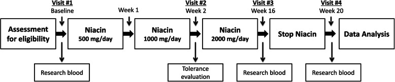
Methods: A 20-week investigational study was performed with 11 participants receiving extended-release niacin (target dose = 2 g/day) for 16-weeks followed by a 4-week washout period. HDL was isolated from participants at weeks: 0, 16, and 20. The HDL proteome was analyzed at each time point by mass spectrometry and relative protein quantification was performed by label-free precursor ion intensity measurement.
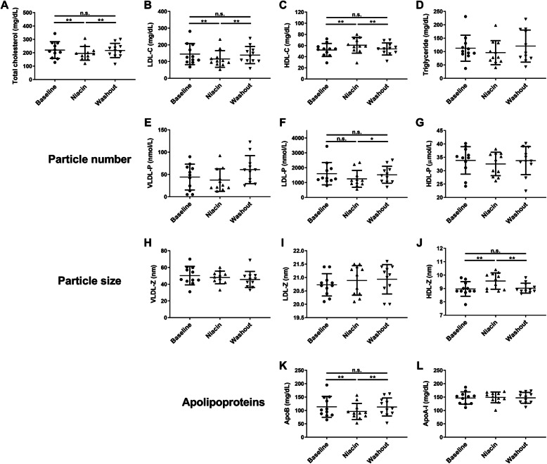
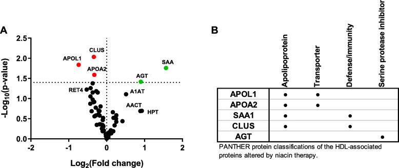
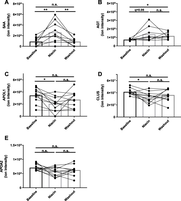
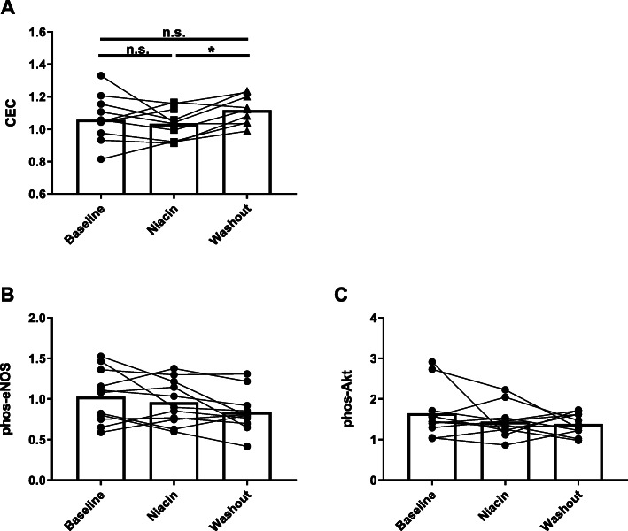
Results: In this cohort, niacin therapy had typical effects on routine clinical lipids (HDL-C + 16%, q < 0.01; LDL-C - 20%, q < 0.01; and triglyceride - 15%, q = 0.1). HDL proteomics revealed significant effects of niacin on 5 proteins: serum amyloid A (SAA), angiotensinogen (AGT), apolipoprotein A-II (APOA2), clusterin (CLUS), and apolipoprotein L1 (APOL1). SAA was the most prominently affected protein, increasing 3-fold in response to niacin (q = 0.008). Cholesterol efflux capacity was not significantly affected by niacin compared to baseline, however, stopping niacin resulted in a 9% increase in efflux (q < 0.05). Niacin did not impact HDL's ability to influence endothelial function.
Conclusion: Extended-release niacin therapy, in the absence of other lipid-modifying medications, can increase HDL-associated SAA, an acute phase protein associated with HDL dysfunction.
Keywords: Apolipoproteins; Cholesterol efflux; High density lipoprotein; Niacin; Proteomics; Serum amyloid a; Vitamin B3.
Conflict of interest statement
The authors declare that they have no competing interests.
Figures
Similar articles
-
Effect of Extended-Release Niacin on High-Density Lipoprotein (HDL) Functionality, Lipoprotein Metabolism, and Mediators of Vascular Inflammation in Statin-Treated Patients.J Am Heart Assoc. 2015 Sep 15;4(9):e001508. doi: 10.1161/JAHA.114.001508. J Am Heart Assoc. 2015. PMID: 26374297 Free PMC article. Clinical Trial.
-
Niacin Increases Atherogenic Proteins in High-Density Lipoprotein of Statin-Treated Subjects.Arterioscler Thromb Vasc Biol. 2021 Aug;41(8):2330-2341. doi: 10.1161/ATVBAHA.121.316278. Epub 2021 Jun 17. Arterioscler Thromb Vasc Biol. 2021. PMID: 34134520 Free PMC article. Clinical Trial.
-
Effects of niacin and omega-3 fatty acids on the apolipoproteins in overweight patients with elevated triglycerides and reduced HDL cholesterol.Atherosclerosis. 2015 Jun;240(2):520-5. doi: 10.1016/j.atherosclerosis.2015.04.793. Epub 2015 Apr 22. Atherosclerosis. 2015. PMID: 25932792 Clinical Trial.
-
Niacin and cholesterol: role in cardiovascular disease (review).J Nutr Biochem. 2003 Jun;14(6):298-305. doi: 10.1016/s0955-2863(02)00284-x. J Nutr Biochem. 2003. PMID: 12873710 Review.
-
New developments in the use of niacin for treatment of hyperlipidemia: new considerations in the use of an old drug.Coron Artery Dis. 1996 Apr;7(4):321-6. doi: 10.1097/00019501-199604000-00009. Coron Artery Dis. 1996. PMID: 8853585 Review.
Cited by
-
Differences in vascular endothelial function and serum proteome between obese people with phlegm-dampness constitution and balanced constitution.J Tradit Chin Med. 2024 Feb;44(1):188-196. doi: 10.19852/j.cnki.jtcm.20231110.005. J Tradit Chin Med. 2024. PMID: 38213254 Free PMC article.
-
Association between dietary niacin intake and dyslipidemia prevalence in the National Health and Nutrition Examination Surveys (NHANES).Asia Pac J Clin Nutr. 2025 Apr;34(2):183-192. doi: 10.6133/apjcn.202504_34(2).0005. Asia Pac J Clin Nutr. 2025. PMID: 40134057 Free PMC article.
-
Association of dietary niacin intake with all-cause mortality in chronic kidney disease: A retrospective cohort study of NHANES.PLoS One. 2025 Feb 7;20(2):e0313398. doi: 10.1371/journal.pone.0313398. eCollection 2025. PLoS One. 2025. PMID: 39919081 Free PMC article.
-
Flaxseed Reduces Cancer Risk by Altering Bioenergetic Pathways in Liver: Connecting SAM Biosynthesis to Cellular Energy.Metabolites. 2023 Aug 14;13(8):945. doi: 10.3390/metabo13080945. Metabolites. 2023. PMID: 37623888 Free PMC article.
-
HDL-associated vitamin D binding protein levels are inversely associated with necrotic plaque burden in psoriasis.Atheroscler Plus. 2024 Dec 13;59:32-38. doi: 10.1016/j.athplu.2024.12.002. eCollection 2025 Mar. Atheroscler Plus. 2024. PMID: 39811778 Free PMC article.
References
MeSH terms
Substances
Grants and funding
LinkOut - more resources
Full Text Sources
Miscellaneous
