. 2021 Mar 1;89(5):486-496.
doi: 10.1016/j.biopsych.2020.06.014. Epub 2020 Jun 23.

Atypical Neurogenesis in Induced Pluripotent Stem Cells From Autistic Individuals

Affiliations

Atypical Neurogenesis in Induced Pluripotent Stem Cells From Autistic Individuals

Dwaipayan Adhya et al. Biol Psychiatry. .

Abstract

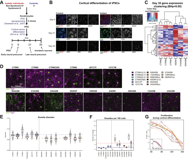
Background: Autism is a heterogeneous collection of disorders with a complex molecular underpinning. Evidence from postmortem brain studies have indicated that early prenatal development may be altered in autism. Induced pluripotent stem cells (iPSCs) generated from individuals with autism with macrocephaly also indicate prenatal development as a critical period for this condition. But little is known about early altered cellular events during prenatal stages in autism.

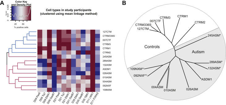
Methods: iPSCs were generated from 9 unrelated individuals with autism without macrocephaly and with heterogeneous genetic backgrounds, and 6 typically developing control individuals. iPSCs were differentiated toward either cortical or midbrain fates. Gene expression and high throughput cellular phenotyping was used to characterize iPSCs at different stages of differentiation.

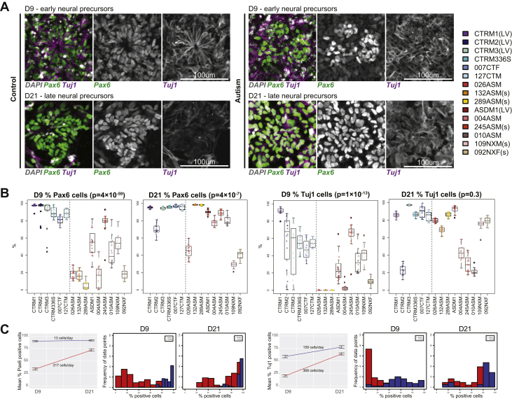
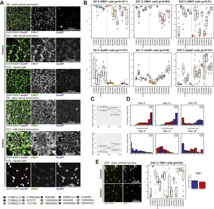
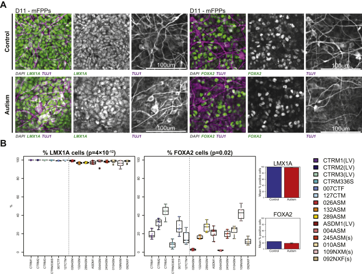
Results: A subset of autism-iPSC cortical neurons were RNA-sequenced to reveal autism-specific signatures similar to postmortem brain studies, indicating a potential common biological mechanism. Autism-iPSCs differentiated toward a cortical fate displayed impairments in the ability to self-form into neural rosettes. In addition, autism-iPSCs demonstrated significant differences in rate of cell type assignment of cortical precursors and dorsal and ventral forebrain precursors. These cellular phenotypes occurred in the absence of alterations in cell proliferation during cortical differentiation, differing from previous studies. Acquisition of cell fate during midbrain differentiation was not different between control- and autism-iPSCs.

Conclusions: Taken together, our data indicate that autism-iPSCs diverge from control-iPSCs at a cellular level during early stage of neurodevelopment. This suggests that unique developmental differences associated with autism may be established at early prenatal stages.

Keywords: Autism; Cortical differentiation; Functional genomics; Midbrain differentiation; Neural precursors; Neural progenitor cells; Neurodevelopment.


Figures

Figure 1
Figure 1
Differentiation of iPSCs into cortical lineage reveals gene expression and neural rosette formation differences between control and autism. (A) Study design and differentiation time points used in this study. (B) Differentiation of control- and autism-iPSCs generate precursor markers Ki67, Nestin, and PAX6 and neuronal markers TBR1 and MAP2. (C) Differential gene expression and hierarchical clustering reveals significant differences between control and autism samples (biological replicates for each sample labeled 1 and 2). (D) Day 9 neural rosette morphology from all participants in this study. (E) Rosette diameter violin plot (horizontal lines show mean rosette diameter; blue, control: red-dashed, autism). (F) Number of rosettes per 100 cells (horizontal lines show mean rosette number; blue, control; red-dashed, autism). (G) Proliferation during cortical differentiation at days 0, 9, 21, and 35 (dashed lines are control samples; color key on top right corner). BH, Benjamini-Hochberg; EdU, 5-ethynyl-2′-deoxyuridine; iPSC, induced pluripotent stem cells; LV, lentivirus reprogramming method used for generating these iPSCs; s, participants with syndromic autism; ZO-1, zonula occludens-1.
Figure 2
Figure 2
Atypical cortical differentiation of autism induced pluripotent stem cells. (A) At day 9 and day 21 precursor cell stages, both control– and autism–induced pluripotent stem cells expressed PAX6 and Tuj1. (B) Quantification of PAX6+ and Tuj1+ cells of individual participants (percentage of cells positive per experimental replicate) showed significant differences between the autism and control samples. (C) Mean values of percentage of positive cells over time show significant difference between control and autism groups at days 9 and 21, as well as significant difference in rate of appearance of markers. Histogram shows normal distribution of experimental data points and demonstrates variability between control and autism samples. D, day; LV, lentivirus reprogramming method used for generating these induced pluripotent stem cells; s, participants with syndromic autism.
Figure 3
Figure 3
Atypical differentiation into dorsal and ventral forebrain precursors in autism. (A) EMX1 was expressed at day 9, day 21, and day 35 in both control and autism groups. GAD67 expression in both groups was time dependent, it decreased over time in in the control sample, while it increased over time in the autism sample. (B) Quantification of EMX1+ and GAD67+ cells (percentage of cells positive per experimental replicate) showed significant differences between autism and control samples. (C) Mean values of percentage of positive cells over time show significant difference between control and autism samples at every time point, except for GAD67 at day 21 of the precursor stage. (D) Histogram shows normal distribution of experimental data points and clear difference in distribution of data points between groups. (E) Control– and autism–induced pluripotent stem cells also expressed TBR1 at day 35 of cortical differentiation, and TBR1 expression was marginally higher in control vs. autism samples. D, day; LV, lentivirus reprogramming method used for generating these induced pluripotent stem cells; s, participants with syndromic autism.
Figure 4
Figure 4
Efficient differentiation of control and autism-iPSCs toward a midbrain fate. (A) Both control- and autism-iPSCs expressed LMX1A and FOXA2 when differentiated into mFPP cells. (B) Differences between control and autism-iPSCs expressing LMX1A or FOXA2 was near negligible. iPSC, induced pluripotent stem cells; LV, lentivirus reprogramming method used for generating these iPSCs; mFPP, midbrain floor plate precursor; s, participants with syndromic autism.
Figure 5
Figure 5
Hierarchical clustering of cellular data using mean linkage method. (A) All control and autism-iPSC lines were grouped based on percentage of positive values for PAX6, Tuj1, EMX1, and GAD67 at day 9, day 21, and day 35 cortical differentiation and LMX1A and FOXA2 at day 11 of midbrain differentiation. Control participants and participants with autism were grouped separately using this unsupervised learning method. (B) Unrooted phylogenetic tree showing relatedness of individual participants based on cellular phenotypes. ∗Syndromic samples branched separately to nonsyndromic samples. ∗NRXN1 deletion samples grouped together on the same branch. 004ASM and 010ASM, which grouped on the same branch (shown with dashed lines), also grouped similarly based on gene expression data shown in Figure 1C and Figure S3B in Supplement 1. D, day; iPSC, induced pluripotent stem cell.

Comment in

References

    1. Berg J.M., Geschwind D.H. Autism genetics: Searching for specificity and convergence. Genome Biol. 2012;13:247. - PMC - PubMed
    1. Bourgeron T. From the genetic architecture to synaptic plasticity in autism spectrum disorder. Nat Rev Neurosci. 2015;16:551–563. - PubMed
    1. O'Roak B.J., Vives L., Girirajan S., Karakoc E., Krumm N., Coe B.P. Sporadic autism exomes reveal a highly interconnected protein network of de novo mutations. Nature. 2012;485:246–250. - PMC - PubMed
    1. Johnson C.P., Myers S.M., American Academy of Pediatrics Council on Children With D Identification and evaluation of children with autism spectrum disorders. Pediatrics. 2007;120:1183–1215. - PubMed
    1. Schubert D., Martens G.J., Kolk S.M. Molecular underpinnings of prefrontal cortex development in rodents provide insights into the etiology of neurodevelopmental disorders. Mol Psychiatry. 2015;20:795–809. - PMC - PubMed

Publication types

LinkOut - more resources