. 2020 Aug 11;11(8):687.
doi: 10.1038/s41419-020-02826-x.

High expression of PTPRM predicts poor prognosis and promotes tumor growth and lymph node metastasis in cervical cancer

Affiliations

High expression of PTPRM predicts poor prognosis and promotes tumor growth and lymph node metastasis in cervical cancer

Pan Liu et al. Cell Death Dis. .

Abstract

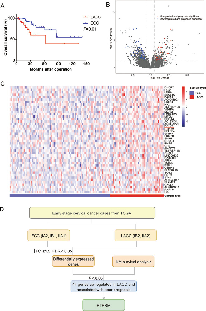
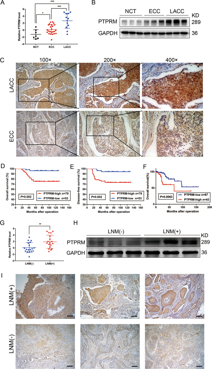
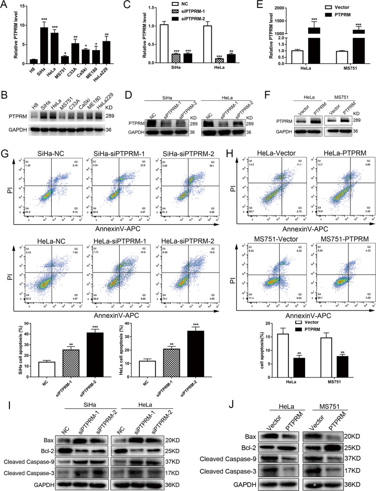
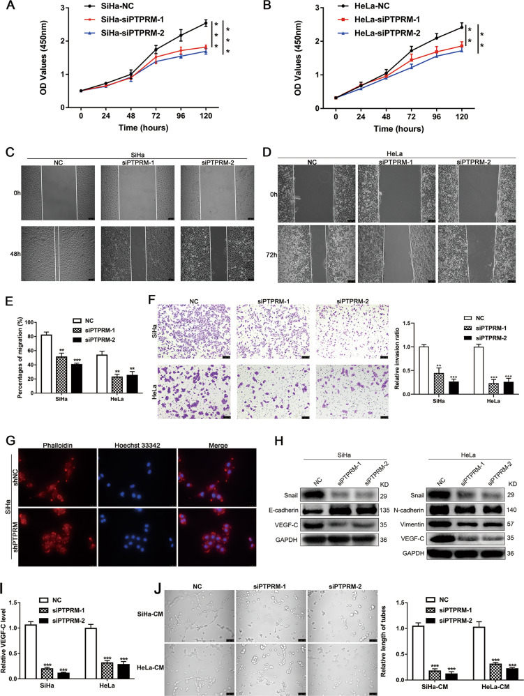
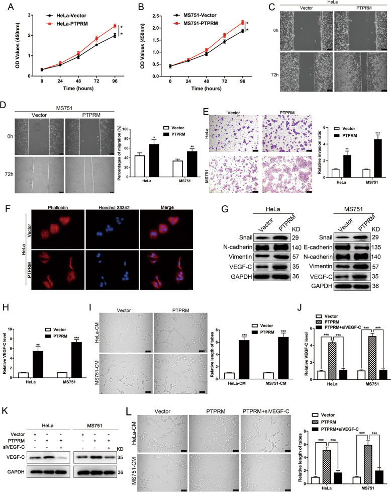
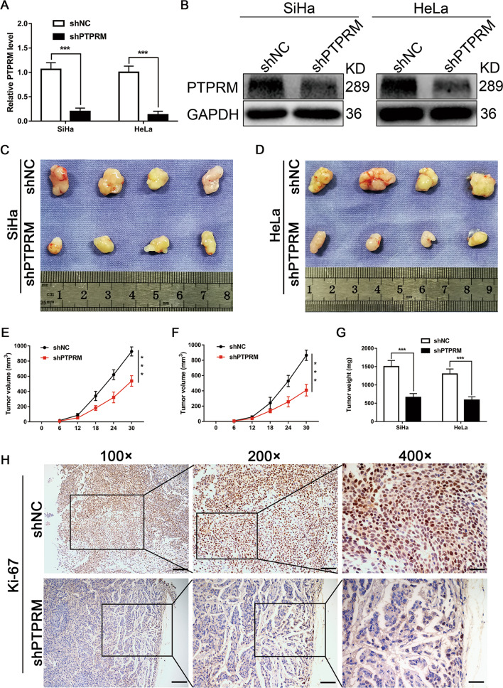
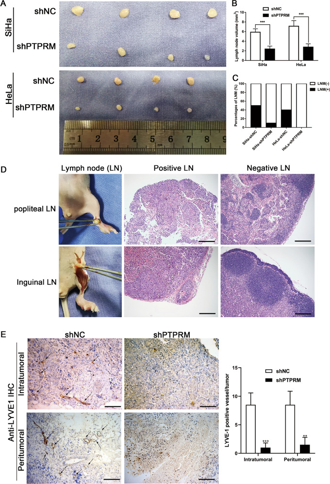
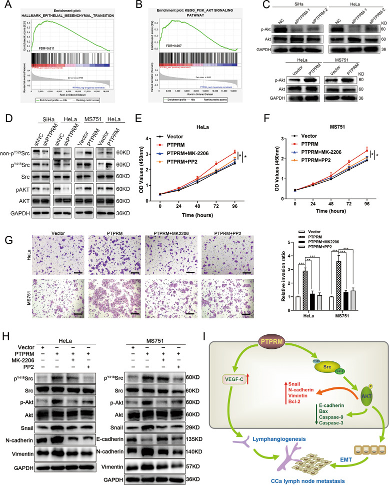
The prognosis for cervical cancer (CCa) patients with lymph node metastasis (LNM) is dismal. Elucidation of the molecular mechanisms underlying LNM may provide clinical therapeutic strategies for CCa patients with LNM. However, the precise mechanism of LNM in CCa remains unclear. Herein, we demonstrated that protein tyrosine phosphatase receptor type M (PTPRM), identified from TCGA dataset, was markedly upregulated in CCa with LNM and correlated with LNM. Moreover, PTPRM was an independent prognostic factor of CCa patients in multivariate Cox's proportional hazards model analysis and associated with poor prognosis. Furthermore, through gain-of-function and loss-of-function approaches, we found that PTPRM promoted CCa cells proliferation, migration, invasion, lymphangiogenesis, and LNM. Mechanistically, PTPRM promoted epithelial-mesenchymal transition (EMT) via Src-AKT signaling pathway and induced lymphangiogenesis in a VEGF-C dependent manner, resulting in LNM of CCa. Importantly, knockdown of PTPRM dramatically reduced LNM in vivo, suggesting that PTPRM plays an important role in the LNM of CCa. Taken together, our findings uncover a novel molecular mechanism in the LNM of CCa and identify PTPRM as a novel prognostic factor and potential therapeutic target for LNM in CCa.


Conflict of interest statement

The authors declare that they have no conflict of interest.

Figures

Fig. 1
Fig. 1. Identification of PTPRM in CCa from TCGA dataset.
a LACC group patients had a worse OS compared with ECC group in TCGA database. b The volcano plot showed the differential expressed genes between LACC and ECC group in TCGA database. c Heat map presented 44 genes highly expressed in LACC group and related to the poor prognosis of CCa. d The detailed screening flowchart for PTPRM.
Fig. 2
Fig. 2. PTPRM is upregulated in CCa with LNM and correlates with LNM and poor prognosis.
a, b The results of qRT-PCR and Western blot showed PTPRM was up-regulated in LACC group patients compared with ECC group and normal cervix tissues (NCT). c Representative IHC images confirmed the expression level of PTPRM in ECC and LACC tissues. Original magnification: ×100, ×200, ×400. d, e CCa patients with high PTPRM level had a poor OS and DFS analyzed in our specimen cohort (IA2-IIA2). f TCGA database showed the expression of PTPRM was negatively associated with OS of CCa patients (IA2-IIA2). g, h qRT-PCR and Western blot demonstrated that PTPRM presented a higher expression in lymphatic metastatic CCa tissues. i The representing IHC images of PTPRM in CCa tissues with or without lymphatic metastasis. Original magnification: ×100. *, ** or ***: significantly different from the corresponding control, P < 0.05, P < 0.01 or P < 0.001, respectively.
Fig. 3
Fig. 3. PTPRM inhibits cell apoptosis in vitro.
a, b PTPRM mRNA and protein expression level in 7 CCa cell lines and normal cervix derived cell line H8. cf qRT-PCR and Western blot confirmed the efficiency of PTPRM knockdown and overexpression. g, h Flow cytometry analysis for Annexin V-APC and PI staining in CCa cells transfected with siRNAs or plasmids for 72 h. i, j Bax, Bcl-2, cleaved caspase-9 and cleaved caspase-3 expression after PTPRM knockdown or overexpression *, ** or ***: significantly different from the corresponding control, P < 0.05, P < 0.01 or P < 0.001, respectively.
Fig. 4
Fig. 4. Downregulation PTPRM inhibits CCa proliferation, invasion, EMT, and lymphangiogenesis in vitro.
a, b CCK8 assays revealed that the proliferation ability of SiHa and HeLa cells was significantly inhibited in the PTPRM knockdown cells. ce Wound-healing assays were performed to investigate PTPRM knockdown suppressed migration of cervical cancer cells. Original magnification: ×100. f Transwell assays showed PTPRM knockdown impeded invasion of cervical cancer cells. Original magnification: ×100. g The morphology change of SiHa cells shown by TRITC phalloidin staining after PTPRM knockdown. Original magnification: ×400. h Western blot analysis of Snail, E-cadherin, N-cadherin, Vimentin and VEGF-C expression level in CCa cells of NC group and PTPRM knockdown group. i qRT-PCR confirmed VEGF-C expression level after PTPRM knockdown in CCa cells. j HLECs tube formation assay demonstrated the effect of PTPRM on lymphangiogenesis. Original magnification: ×200. Average length of tube formation was quantified. *, ** or ***: significantly different from the corresponding control, P < 0.05, P < 0.01 or P < 0.001, respectively.
Fig. 5
Fig. 5. PTPRM overexpression promotes CCa proliferation, invasion, EMT, and lymphangiogenesis in vitro.
a, b CCK8 assay showed PTPRM overexpression promoted the proliferation of CCa cells. ce Wound healing and transwell assays indicated that PTPRM overexpression increased the migration and invasion ability of CCa cells. Original magnification: ×100. f The morphology change of HeLa cells shown by TRITC phalloidin staining after PTPRM overexpression. Original magnification: ×400. g Snail, E-cadherin, N-cadherin, Vimentin and VEGF-C expression level were confirmed by Western blot in PTPRM overexpression cells. h qRT-PCR confirmed VEGF-C expression level after PTPRM expression in CCa cells. i PTPRM overexpression promoted HLECs tube formation. jk VEGF-C mRNA and protein levels after PTPRM overexpressed cells were treated with siVEGF-C. l VEGF-C knockdown abrogated PTPRM overexpression induced lymphangiogenesis. Total length of tube formation was quantified. Original magnification: ×200. *, ** or ***: significantly different from the corresponding control, P < 0.05, P < 0.01 or P < 0.001, respectively.
Fig. 6
Fig. 6. PTPRM promotes tumor growth of CCa in vivo.
a, b qRT-PCR and Western blot were used to confirm the efficiency of transduction shPTPRM in SiHa and HeLa cells. c, d The representative pictures of subcutaneous tumors from nude mice injected cells for 30 days (n = 10 per group). eg Subcutaneous tumor volume and weight of mice in different treatment groups were displayed. h The Ki-67 expression levels of subcutaneous tumors from shNC and shPTPRM groups mice. Original magnification, ×100, ×200, ×400. *, ** or ***: significantly different from the corresponding control, P < 0.05, P < 0.01 or P < 0.001, respectively.
Fig. 7
Fig. 7. PTPRM knockdown suppresses the lymphangiogenesis and LNM of CCa in vivo.
a The representative pictures of popliteal lymph nodes (n = 10 per group). b, c Histogram analysis of the average popliteal lymph nodes volume and the rate of LNM in different treatment groups. d The representative pictures of H-E staining of positive and negative metastatic popliteal lymph nodes and inguinal lymph nodes. Original magnification: ×100. e Representative IHC pictures of intratumoral and peritumoral lymphatic vessels in the primary tumors resected from footpads of nude mice stained by anti-LYVE-1 in the mice bearing PTPRM-silenced cells and control group (left panel), quantification of LYVE-1 positive vessels in primary tumors (right panel). Original magnification: ×200. *, ** or ***: significantly different from the corresponding control, P < 0.05, P < 0.01 or P < 0.001, respectively.
Fig. 8
Fig. 8. PTPRM promotes CCa proliferation and metastasis through Src-AKT signaling pathway.
a, b GSEA showed PTPRM was positively correlated with AKT and EMT pathway. c Western blot analysis revealed p-AKT level in CCa cells with PTPRM knockdown or overexpression. d Western blot analysis revealed non-pY529Src, pY418Src, and p-AKT levels in CCa cells with PTPRM knockdown or overexpression. e, f CCK8 assay suggested that PP2 and MK-2206 abrogated the promoting effect of PTPRM upregulation on cell proliferation ability in HeLa and MS751 cells. g Transwell assay demonstrated that PP2 and MK-2206 alleviated the increased invasion capability due to PTPRM overexpression in HeLa and MS751 cells. h Western blot analysis showed that pY418Src, p-AKT, Snail, E-cadherin, N-cadherin, Vimentin expression levels were partly rescued by PP2 and MK-2206 treatment in CCa cells. i Schematic illustration showing the proposed mechanism by which PTPRM promotes CCa lymph node metastasis. *, ** or ***: significantly different from the corresponding control, P < 0.05, P < 0.01 or P < 0.001, respectively.

Similar articles

Cited by

References

    1. Bray F, et al. Global cancer statistics 2018: GLOBOCAN estimates of incidence and mortality worldwide for 36 cancers in 185 countries. CA Cancer J. Clin. 2018;68:394–424. doi: 10.3322/caac.21492. - DOI - PubMed
    1. Cohen PA, Jhingran A, Oaknin A, Denny L. Cervical cancer. Lancet. 2019;393:169–182. doi: 10.1016/S0140-6736(18)32470-X. - DOI - PubMed
    1. Van Trappen PO, et al. Molecular quantification and mapping of lymph-node micrometastases in cervical cancer. Lancet. 2001;357:15–20. doi: 10.1016/S0140-6736(00)03566-2. - DOI - PubMed
    1. Hou T, et al. Atypical chemokine receptors predict lymph node metastasis and prognosis in patients with cervical squamous cell cancer. Gynecol. Oncol. 2013;130:181–187. doi: 10.1016/j.ygyno.2013.04.015. - DOI - PubMed
    1. Niu, C. et al. NR2F6 expression correlates with pelvic lymph node metastasis and poor prognosis in early-stage cervical cancer. Int. J. Mol. Sci.17, 1694 (2016). - PMC - PubMed

Publication types

MeSH terms

Substances