. 2020 Sep 10;530(1):107-114.
doi: 10.1016/j.bbrc.2020.07.031. Epub 2020 Jul 30.

AAV9-DOK7 gene therapy reduces disease severity in Smn2B/- SMA model mice

Affiliations

AAV9-DOK7 gene therapy reduces disease severity in Smn2B/- SMA model mice

Kevin A Kaifer et al. Biochem Biophys Res Commun. .

Abstract

Spinal Muscular Atrophy (SMA) is an autosomal recessive neuromuscular disease caused by deletions or mutations in the survival motor neuron (SMN1) gene. An important hallmark of disease progression is the pathology of neuromuscular junctions (NMJs). Affected NMJs in the SMA context exhibit delayed maturation, impaired synaptic transmission, and loss of contact between motor neurons and skeletal muscle. Protection and maintenance of NMJs remains a focal point of therapeutic strategies to treat SMA, and the recent implication of the NMJ-organizer Agrin in SMA pathology suggests additional NMJ organizing molecules may contribute. DOK7 is an NMJ organizer that functions downstream of Agrin. The potential of DOK7 as a putative therapeutic target was demonstrated by adeno-associated virus (AAV)-mediated gene therapy delivery of DOK7 in Amyotrophic Lateral Sclerosis (ALS) and Emery Dreyefuss Muscular Dystrophy (EDMD). To assess the potential of DOK7 as a disease modifier of SMA, we administered AAV-DOK7 to an intermediate mouse model of SMA. AAV9-DOK7 treatment conferred improvements in NMJ architecture and reduced muscle fiber atrophy. Additionally, these improvements resulted in a subtle reduction in phenotypic severity, evidenced by improved grip strength and an extension in survival. These findings reveal DOK7 is a novel modifier of SMA.

Keywords: Adeno associate virus; Gene Therapy; Neurodegeneration; SMA; Spinal muscular atrophy.


Conflict of interest statement

Declaration of competing interest C.L.L. is the co-founder and Chief Scientific Officer of Shift Pharmaceuticals.

Figures

Figure 1:
Figure 1:. scAAV9-DOK7 improves phenotype of Smn2B/− mice.
(A) Western blot of DOK7 in hindlimb skeletal muscle. (B) Western blot of SMN in hindlimb skeletal muscle. (C) Kaplan-Meier curve of Smn2B/− mice treated with scAAV9-DOK7. Difference between scAAV9-DOK7-treated and untreated Smn2B/− mice was calculated using log-rank Mantel-Cox test (D) Weight gain of Smn2B/− mice treated with scAAV9-DOK7. (E) Average grip strength on P17 and P21. Groups were compared using Student’s t-test. (*P<0.05, ***P<0.001)
Figure 2:
Figure 2:. scAAV9-DOK7 improves endplate area of NMJs in Smn2B/− mice.
Transverse abdominus muscles were dissected from mice harvested on P17. (A) Representative images of neuromuscular junction endplates labelled with α-bungarotoxin (red). Images were taken with 40X objective lens (scale bars = 20 μm). (B) Scatter plot representing quantification of motor endplate area. Bars represent mean ± SEM. (C) Representative images of neuromuscular junctions stained with neurofilament (green) and synaptophysin (green) to label the presynaptic terminals and α-bungarotoxin (red) to label endplates. (D) Bar graph representing average percentage of fully innervated, partially innervated, and denervated motor endplates. Bars represent mean ± SEM. Statistical analysis was performed comparing degree of fully-innervated endplates. Experimental groups were compared using one-way ANOVA with Tukey’s multiple comparisons test. (Smn2B/+ n=4; Smn2B/− n=7; Smn2B/− + scAAV9-DOK7 n=7) (***P < 0.001, nsP > 0.05)
Figure 3:
Figure 3:. scAAV9-DOK7 does not improve central defects in Smn2B/− mice.
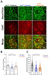
(A) Motor neuron soma analyses were performed at P21 (n=3 mice). Representative images of immunofluorescent labelling of ChAT-positive (red) motor neuron soma and NeuroTrace Nissl (green) in the L4-L6 spinal cord (Scale bars = 50 μm). (B) Scatter plot representing number of ChAT-positive motor neuron soma in the L4-L6 spinal cord. (C) Scatter plot representing cross sectional area of ChAT-positive motor neuron soma in the L4-L6 spinal cord. Bars represent mean ± SEM. Experimental groups were compared using one-way ANOVA with Tukey’s multiple comparisons test. (**P < 0.01, ***P < 0.001, nsP > 0.05)
Figure 4:
Figure 4:. scAAV9-DOK7 reduces muscle fiber pathology in Smn2B/− mice.
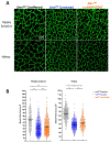
Muscle analysis was performed at P17 (n=4 mice). (A) Representative images of cross sections of tibialis anterior and soleus muscles dissected on P17. Sections were stained with laminin (green). Images were taken with 40× objective lens (Scale bars = 50 μm). (B) Scatter plot representing cross sectional area of TA and SO muscle fibers. Experimental groups were compared using one-way ANOVA with Tukey’s multiple comparisons test. (***P < 0.001)

References

    1. Sugarman EA et al. Pan-ethnic carrier screening and prenatal diagnosis for spinal muscular atrophy: clinical laboratory analysis of >72,400 specimens. European journal of human genetics : EJHG 20, 27–32, doi:10.1038/ejhg.2011.134 (2012). - DOI - PMC - PubMed
    1. Crawford TO & Pardo CA The neurobiology of childhood spinal muscular atrophy. Neurobiology of disease 3, 97–110, doi:10.1006/nbdi.1996.0010 (1996). - DOI - PubMed
    1. Coady TH & Lorson CL SMN in spinal muscular atrophy and snRNP biogenesis. Wiley interdisciplinary reviews. RNA 2, 546–564, doi:10.1002/wrna.76 (2011). - DOI - PubMed
    1. Fallini C, Bassell GJ & Rossoll W Spinal muscular atrophy: the role of SMN in axonal mRNA regulation. Brain research 1462, 81–92, doi:10.1016/j.brainres.2012.01.044 (2012). - DOI - PMC - PubMed
    1. Hoy SM Nusinersen: A Review in 5q Spinal Muscular Atrophy. CNS Drugs 32, 689–696, doi:10.1007/s40263-018-0545-1 (2018). - DOI - PubMed

Publication types

MeSH terms