

Role of histone deacetylases in bone development and skeletal disorders
- PMID: 32829038
- PMCID: PMC7770092
- DOI: 10.1016/j.bone.2020.115606
Role of histone deacetylases in bone development and skeletal disorders
Abstract
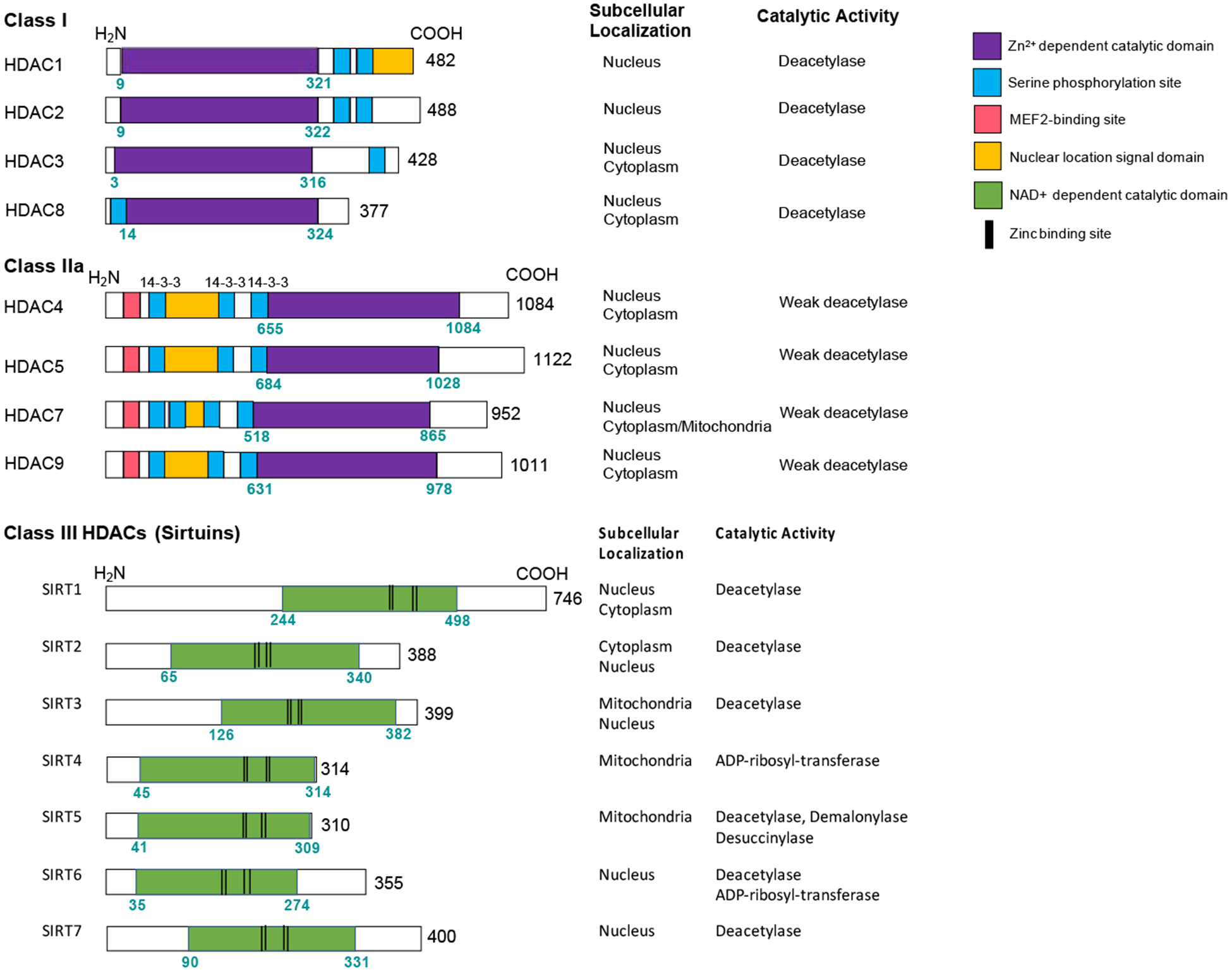
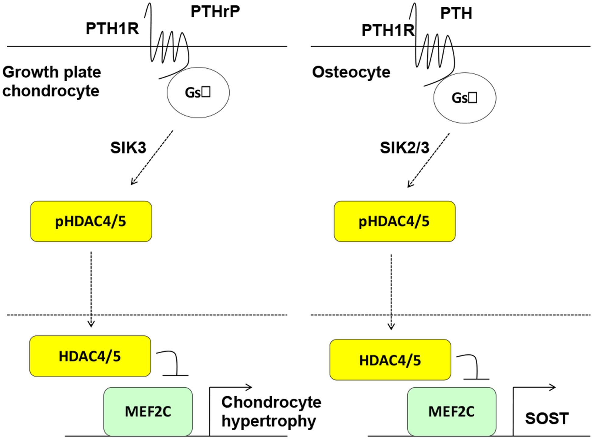
Bone cells must constantly respond to hormonal and mechanical cues to change gene expression programs. Of the myriad of epigenomic mechanisms used by cells to dynamically alter cell type-specific gene expression, histone acetylation and deacetylation has received intense focus over the past two decades. Histone deacetylases (HDACs) represent a large family of proteins with a conserved deacetylase domain first described to deacetylate lysine residues on histone tails. It is now appreciated that multiple classes of HDACs exist, some of which are clearly misnamed in that acetylated lysine residues on histone tails is not the major function of their deacetylase domain. Here, we will review the roles of proteins bearing deacetylase domains in bone cells, focusing on current genetic evidence for each individual HDAC gene. While class I HDACs are nuclear proteins whose primary role is to deacetylate histones, class IIa and class III HDACs serve other important cellular functions. Detailed knowledge of the roles of individual HDACs in bone development and remodeling will set the stage for future efforts to specifically target individual HDAC family members in the treatment of skeletal diseases such as osteoporosis.
Keywords: Bone development; Histone deacetylase; Salt inducible kinase; Sirtuin.
Copyright © 2020 Elsevier Inc. All rights reserved.
Conflict of interest statement
Competing Interests Statement
MNW receives research funding from Radius Health and Galapagos NV on projects unrelated to this review. These funders had no role in preparation of this review manuscript or decision to publish. All other authors declare no conflict of interest.
Figures
Similar articles
-
Histone deacetylase (HDACs) inhibitors: Clinical applications.Prog Mol Biol Transl Sci. 2023;198:119-152. doi: 10.1016/bs.pmbts.2023.02.011. Epub 2023 Mar 31. Prog Mol Biol Transl Sci. 2023. PMID: 37225317
-
The nonepigenetic role for small molecule histone deacetylase inhibitors in the regulation of cardiac function.Future Med Chem. 2019 Jun;11(11):1345-1356. doi: 10.4155/fmc-2018-0311. Epub 2019 Jun 4. Future Med Chem. 2019. PMID: 31161804 Free PMC article. Review.
-
NAD+-dependent deacetylation of H4 lysine 16 by class III HDACs.Oncogene. 2007 Aug 13;26(37):5505-20. doi: 10.1038/sj.onc.1210617. Oncogene. 2007. PMID: 17694090 Review.
-
Chemical and structural biology of protein lysine deacetylases.Proc Jpn Acad Ser B Phys Biol Sci. 2017;93(5):297-321. doi: 10.2183/pjab.93.019. Proc Jpn Acad Ser B Phys Biol Sci. 2017. PMID: 28496053 Free PMC article. Review.
-
The emerging roles of HDACs and their therapeutic implications in cancer.Eur J Pharmacol. 2022 Sep 15;931:175216. doi: 10.1016/j.ejphar.2022.175216. Epub 2022 Aug 18. Eur J Pharmacol. 2022. PMID: 35988787 Review.
Cited by
-
Hdac1 and Hdac2 positively regulate Notch1 gain-of-function pathogenic signaling in committed osteoblasts of male mice.Birth Defects Res. 2024 Jan;116(1):e2266. doi: 10.1002/bdr2.2266. Epub 2023 Nov 3. Birth Defects Res. 2024. PMID: 37921375 Free PMC article.
-
The Role of Epigenomics in Osteoporosis and Osteoporotic Vertebral Fracture.Int J Mol Sci. 2020 Dec 11;21(24):9455. doi: 10.3390/ijms21249455. Int J Mol Sci. 2020. PMID: 33322579 Free PMC article. Review.
-
Unveiling the signal valve specifically tuning the TGF-β1 suppression of osteogenesis: mediation through a SMAD1-SMAD2 complex.Cell Commun Signal. 2025 Jan 22;23(1):38. doi: 10.1186/s12964-025-02051-z. Cell Commun Signal. 2025. PMID: 39844165 Free PMC article.
-
Urine-derived stem cells-extracellular vesicles ameliorate diabetic osteoporosis through HDAC4/HIF-1α/VEGFA axis by delivering microRNA-26a-5p.Cell Biol Toxicol. 2023 Oct;39(5):2243-2257. doi: 10.1007/s10565-022-09713-5. Epub 2022 May 13. Cell Biol Toxicol. 2023. PMID: 35554780
-
Epigenetic Regulation of Bone Healing: Implications for Fracture Repair and Clinical Treatment Strategies.Yale J Biol Med. 2025 Jun 30;98(2):159-170. doi: 10.59249/HSYL8000. eCollection 2025 Jun. Yale J Biol Med. 2025. PMID: 40589933 Free PMC article. Review.
References
-
- Cavalli G and Heard E, Advances in epigenetics link genetics to the environment and disease. Nature, 2019. 571(7766): p. 489–499. - PubMed
-
- Kornberg RD and Thomas JO, Chromatin structure; oligomers of the histones. Science, 1974. 184(4139): p. 865–8. - PubMed
-
- Luger K, et al., Crystal structure of the nucleosome core particle at 2.8 A resolution. Nature, 1997. 389(6648): p. 251–60. - PubMed
-
- Kouzarides T, Chromatin modifications and their function. Cell, 2007. 128(4): p. 693–705. - PubMed
-
- Zhou VW, Goren A, and Bernstein BE, Charting histone modifications and the functional organization of mammalian genomes. Nat Rev Genet, 2011. 12(1): p. 7–18. - PubMed
Publication types
MeSH terms
Substances
Grants and funding
LinkOut - more resources
Full Text Sources
Other Literature Sources
Molecular Biology Databases
Miscellaneous
