. 2020 Oct;27(10):1373-1378.
doi: 10.1016/j.acra.2020.08.009. Epub 2020 Aug 15.

Thorax Magnetic Resonance Imaging Findings in Patients with Coronavirus Disease (COVID-19)

Affiliations

Thorax Magnetic Resonance Imaging Findings in Patients with Coronavirus Disease (COVID-19)

Omer Faruk Ates et al. Acad Radiol. 2020 Oct.

Abstract

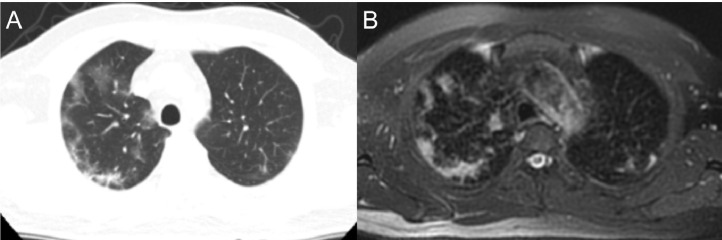
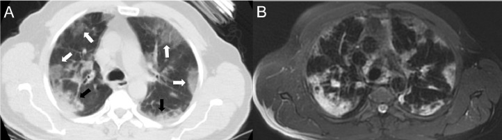
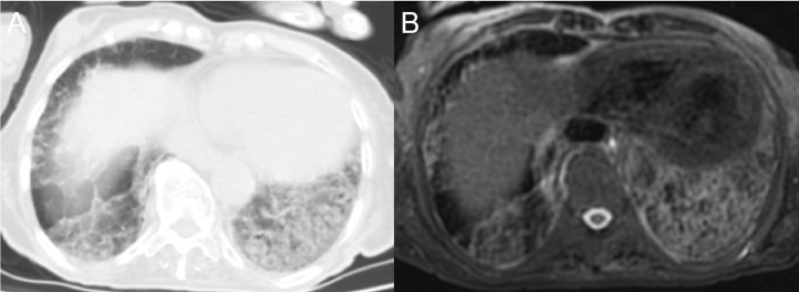
Rationale and objectives: The aim of this study was to compare the findings found in thorax computed tomography (CT), which is increasingly used in the diagnosis of the important public health problem of coronavirus disease (COVID-19), and the findings of magnetic resonance imaging (MRI) as an important diagnostic alternative.

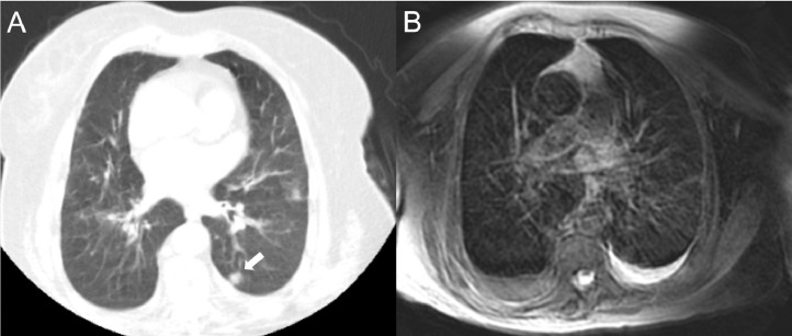
Materials and methods: Thirty-two patients diagnosed with COVID-19 who underwent thorax CT for COVID pneumonia and MRI for any reason within 24 hours after CT were included in the study. The number of lobes affected, number of lobes containing ground-glass opacities and consolidation, number of nodules, distribution of lesions (central, peripheral, or diffuse), lobes with centrilobular nodular pattern, and the presence of pleural effusion were recorded separately for both imaging methods.

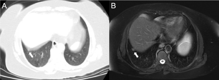
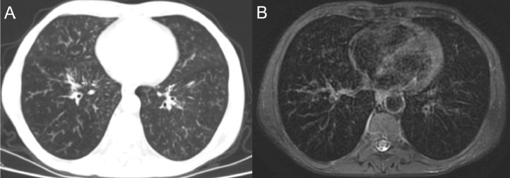
Results: Seventeen of the patients were female (53%) and 15 were male (47%). The mean age of the patients was 60.5 (range, 20-85) years. A total of 31 patients (96%) had signs of pneumonia on CT. The most common finding in CT was ground-glass opacities in 29 patients (90.6%), followed by consolidation in 14 patients (43.75%). Both consolidation and ground-glass opacities were also observed in MRI in all of these patients. Nodules were detected in 12 patients (37.5%) on CT and 11 patients (34.4%) on MRI. The sensitivity and specificity of MRI in nodule detection were calculated as 91.67% and 100%, respectively.

Conclusion: Although thorax CT is widely used in the imaging of COVID-19 infection, due to its advantages, MRI can also be used as an alternative diagnostic tool.

Keywords: COVID-19; Computed tomography; Magnetic resonance imaging.


Figures

Fig. 1
Fig. 1
Images of a 50-year-old male, showing ground-glass areas with bilateral peripheral distribution similarly visualized on both computed tomography (a) and magnetic resonance imaging (b).
Fig. 2
Fig. 2
Images of an 81-year-old woman with peripheral-weighted diffuse involvement. In areas where there are ground-glass appearances (white arrows) on both CT (a) and MRI (b), vascular structures can be selected, while in the consolidated areas, vascular structures cannot be distinguished (black arrows).
Fig. 3
Fig. 3
Crazy-paving pattern in an 81-year-old male patient. Interlobular and intralobular septal thickening and ground-glass appearance are displayed very successfully by MRI (b), as well as CT (a).
Fig. 4
Fig. 4
An 82-year-old female patient with heart failure and chronic obstructive pulmonary disease, presenting with a nodule in the superior segment of the left lung lower lobe displayed clearly on CT (a, arrow), but with poorer quality on MRI (b) due to intense artifacts.
Fig. 5
Fig. 5
A 59-year-old female patient presenting with a nodule (arrow) that is considerably affected by a motion artifact due to its adjacency to the diaphragm in CT examination (a) but can be easily seen on the T2 FSE PROPELLER MRI image due to respiratory gating (b).
Fig. 6
Fig. 6
A 61-year-old male patient with a centrilobular nodular pattern, which is one of the rare findings in COVID-19-related pneumonia. Although the nodules are easier to distinguish on CT, this pattern is also shown very successfully on the MRI image.

References

    1. Zhu N, Zhang D, Wang W. A novel coronavirus from patients with pneumonia in China, 2019. N Engl J Med. 2020;382:727–733. - PMC - PubMed
    1. COVID C, Team R. Severe outcomes among patients with coronavirus disease 2019 (COVID-19)—United States, February 12–March 16, 2020. MMWR Morb Mortal Wkly Rep. 2020;69:343–346. - PMC - PubMed
    1. Xu X, Chen P, Wang J. Evolution of the novel coronavirus from the ongoing Wuhan outbreak and modeling of its spike protein for risk of human transmission. Sci China Life Sci. 2020;63:457–460. - PMC - PubMed
    1. Akcay MS. Radiological approaches to Covid-19 pneumonia. Turk J Med Sci. 2020 - PMC - PubMed
    1. Huang C, Wang Y, Li X. Clinical features of patients infected with 2019 novel coronavirus in Wuhan, China. Lancet. 2020;395:497–506. - PMC - PubMed