. 2020 Dec 22;66(6):515-522.
doi: 10.1262/jrd.2020-082. Epub 2020 Aug 21.

Expression and functions of cluster of differentiation 9 and 81 in rat mammary epithelial cells

Affiliations

Expression and functions of cluster of differentiation 9 and 81 in rat mammary epithelial cells

Kotaro Horiguchi et al. J Reprod Dev. .

Abstract

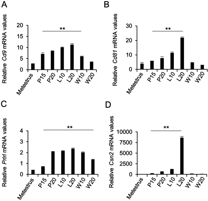
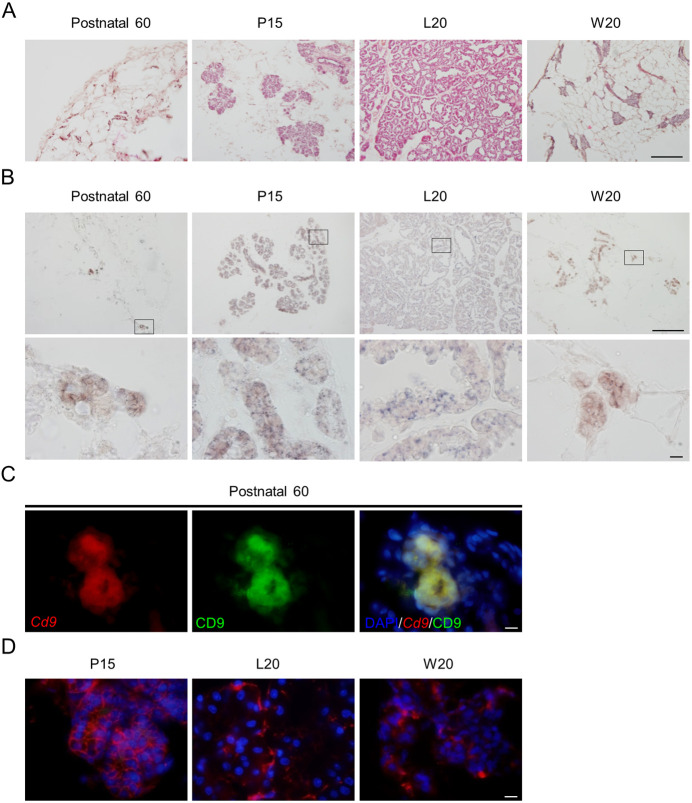
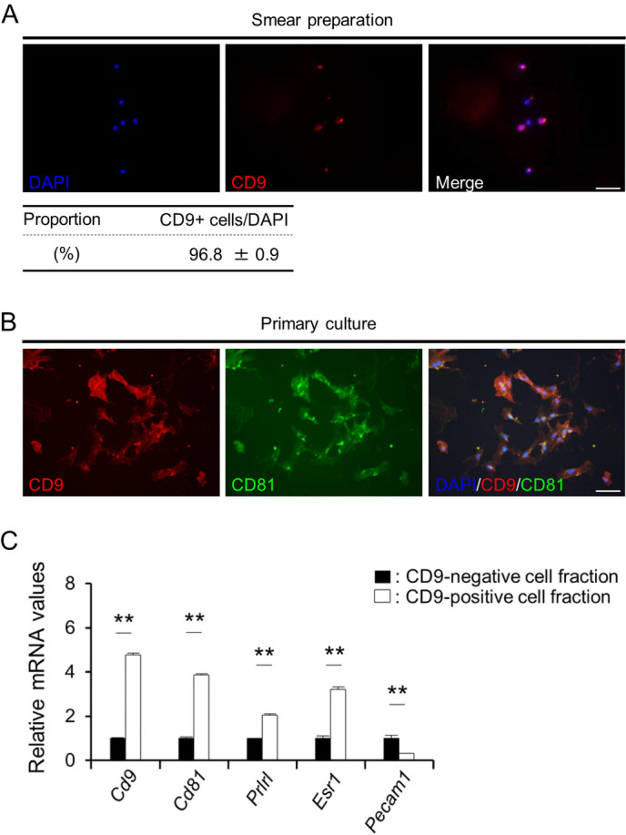
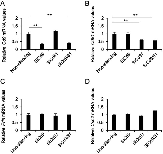
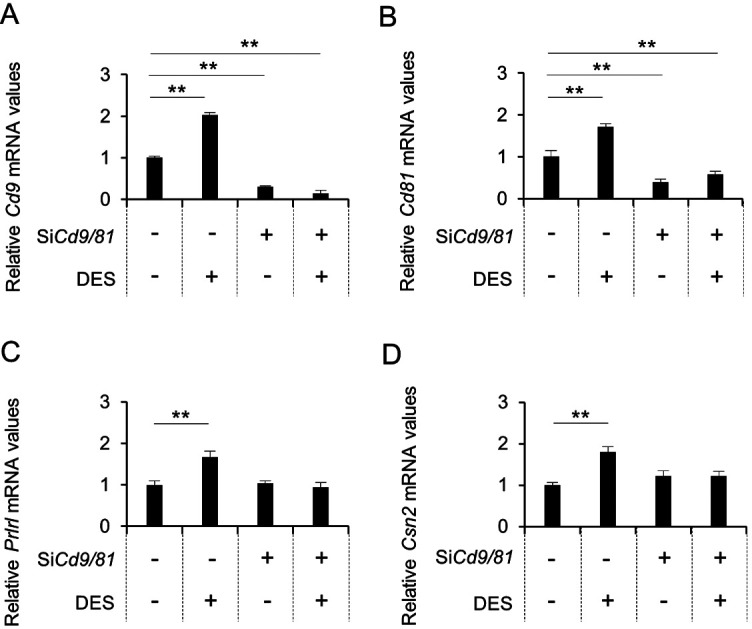
Cluster of differentiation (CD) 9 and CD81 are closely-related members of the tetraspanin family that consist of four-transmembrane domain proteins. Cd9 and Cd81 are highly expressed in breast cancer cells; however, their expression in healthy mammary glands is unclear. In this study, we performed quantitative real-time PCR to analyze the expression levels of Cd9 and Cd81. Histological techniques were employed to identify Cd9- and Cd81-expressing cells in rat mammary glands during pregnancy and lactation. It was observed that Cd9 and Cd81 were expressed in the mammary glands, and their expression levels correlated with mammary gland development. To identify cells expressing Cd9 and Cd81 in the mammary glands, we performed double immunohistochemical staining for CD9 and CD81, prolactin receptor long form, estrogen receptor alpha, or Ki67. The results showed that CD9 and CD81 were co-expressed in proliferating mammary epithelial cells. Next, we attempted to isolate CD9-positive epithelial cells from the mammary gland using pluriBead cell-separation technology based on antibody-mediated binding of cells to beads of different sizes, followed by isolation using sieves with different mesh sizes. We successfully isolated CD9-positive epithelial cells with 96.8% purity. In addition, we observed that small-interfering RNAs against Cd9 and Cd81 inhibited estrogen-induced proliferation of CD9-positive mammary epithelial cells. Our current findings may provide novel insights into the proliferation of mammary epithelial cells during pregnancy and lactation as well as in pathological processes associated with breast cancer.

Keywords: CD81; Cluster of differentiation (CD) 9; Lactation; Mammary gland; Proliferation.


Figures

Fig. 1.
Fig. 1.
Expression of Cd9 and Cd81 in the mammary glands of female rats during pregnancy and lactation. (A) Cd9, (B) Cd81, (C) prolactin receptor long form (Prlrl), and (D) β-casein (Csn2) mRNA expression levels in the mammary glands at metestrus, pregnancy day 15 (P15), P20, lactation day 10 (L10), L20, weaning day 10 (W10), and W20 were determined using real-time PCR (mean ± SEM, n = 3), followed by normalization to the expression of an internal control (Hprt1). ** P < 0.01.
Fig. 2.
Fig. 2.
Immunohistochemistry of CD9-positive cells in the mammary glands during pregnancy and lactation. (A) Hematoxylin and eosin staining of a cryosection of female rat mammary gland at postnatal day 60 (Postnatal 60), pregnancy day 15 (P15), lactation day 20 (L20), and weaning day 20 (W20). (B) In situ hybridization of Cd9 at Postnatal 60, P15, L20, and W20 (upper panels). High-magnification images (lower panels) correspond to the boxed regions shown in the upper panels. (C) In situ hybridization of Cd9 (red, left panel) and immunohistochemistry of CD9 (green, middle panel) at Postnatal 60. Merged images of them and DAPI staining (right panel). (D) Merged images of DAPI staining (blue) and immunofluorescence staining for CD9 (red) at P15, L20, or W20. Scale bars: 100 μm (upper panels of A and B) or 10 μm (lower panel of B; panels of C and D).
Fig. 3.
Fig. 3.
Characterization of CD9-positive cells in the mammary glands at L10. (A) Double immunofluorescence staining of CD9 (left panels) and CD81, ERα, PRLR, or CD133 (top 4 middle panels). In situ hybridization of Cd9 (left panel) and immunofluorescence staining of Ki67 (bottom middle panel). The right panels show merged images of the left and middle panels, together with DAPI staining. Scale bar, 10 μm. White line arrowheads indicate double-positive cells. White arrows indicate CD9-negative cells. (B) Population of CD9-positive (CD9+) cells and cells positive for CD81 (CD81+), ERα (ERα+), PRLR (PRLR+), and CD133 (CD133+), or Cd9-expressing (Cd9+) cells and Ki67-positive (Ki67+) cells in the mammary gland at L10.
Fig. 4.
Fig. 4.
Isolation of CD9-positive cells from female rat mammary glands at L30. (A) DAPI staining (blue) and immunofluorescence staining of CD9 (red) in a smear of CD9-positive cell fraction. The proportion of CD9-positive (CD9+) cells in the CD9-positive cell fraction obtained using pluriBeads are indicated (mean ± SEM, n = 3). The numbers of DAPI-positive and immunopositive cells were counted in random areas of the smear, and the proportion of CD9-positive cells was calculated. (B) Immunofluorescence staining of CD9 in the CD9-positive cell fraction cultured for 72 h. (C) The ratio of the mRNA expression levels of Cd9, Cd81, Esr1, Prlrl, and Pecam1 in CD9-positive cells (white bars) against those in CD9-negative cells (black bars) was determined using qPCR (mean ± SEM, n = 6), followed by normalization to an internal control (Hprt1). The graph shows the relative expression levels of Cd9, Cd81, Prlrl, Esr1, and Pecam1. ** P < 0.01. Scale bars, 50 μm.
Fig. 5.
Fig. 5.
Downregulation of Cd9, Cd81, Prlrl, and Csn2 mRNA levels after transfection of CD9-positive mammary epithelial cells with the indicated siRNAs. (A) Cd9, (B) Cd81, (C) Prlrl, and (D) Csn2 mRNA levels in CD9-positive cells transfected with non-silencing siRNAs, Cd9-siRNA, Cd81-siRNA, or Cd9-siRNA plus Cd81-siRNA for 48 h, as determined using qPCR (mean ± SEM, n = 3), followed by normalization to an internal control (Hprt1). ** P < 0.01.
Fig. 6.
Fig. 6.
Downregulation of Cd9 and Cd81 mRNA levels after transfection of CD9-positive mammary cells with siRNAs and DES treatment. (A) Cd9, (B) Cd81, (C) Prlrl, and (D) Csn2 mRNA expression levels in CD9-positive cells cultured with non-silencing siRNAs or Cd9- and Cd81-siRNAs (black bars) for 48 h, with or without DES, as determined using qPCR (mean ± SEM, n = 3), followed by normalization to an internal control (Hprt1). ** P < 0.01.
Fig. 7.
Fig. 7.
The number of BrdU-positive cells among purified CD9-positive cells treated with DES. (A) Merged images showing DAPI staining (blue), BrdU immunocytochemistry (green), and CD9 immunocytochemistry (red) 48 h after CD9-positive cells were cultured with non-silencing siRNAs (left) or Cd9- and Cd81-siRNAs (right), with or without DES (as indicated). Arrowheads indicate cells that were double-positive for CD9 and BrdU. Scale bar, 10 μm. (B) Proportion of BrdU-positive cells among CD9-positive cells (mean ± SEM, n = 3). ** P < 0.01.

References

    1. Berditchevski F, Odintsova E. Tetraspanins as regulators of protein trafficking. Traffic 2007; 8: 89–96. - PubMed
    1. Brosseau C, Colas L, Magnan A, Brouard S. CD9 Tetraspanin: a new pathway for the regulation of inflammation? Front Immunol 2018; 9: 2316. - PMC - PubMed
    1. Ikeyama S, Koyama M, Yamaoko M, Sasada R, Miyake M. Suppression of cell motility and metastasis by transfection with human motility-related protein (MRP-1/CD9) DNA. J Exp Med 1993; 177: 1231–1237. - PMC - PubMed
    1. Miyake M, Koyama M, Seno M, Ikeyama S. Identification of the motility-related protein (MRP-1), recognized by monoclonal antibody M31-15, which inhibits cell motility. J Exp Med 1991; 174: 1347–1354. - PMC - PubMed
    1. Wang HX, Li Q, Sharma C, Knoblich K, Hemler ME. Tetraspanin protein contributions to cancer. Biochem Soc Trans 2011; 39: 547–552. - PubMed