Transcriptomic Approach for Understanding the Adaptation of Salmonella enterica to Contaminated Produce
- PMID: 32830190
- PMCID: PMC9728351
- DOI: 10.4014/jmb.2007.07036
Transcriptomic Approach for Understanding the Adaptation of Salmonella enterica to Contaminated Produce
Abstract
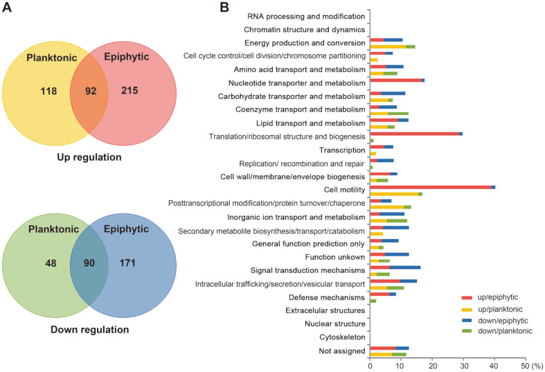
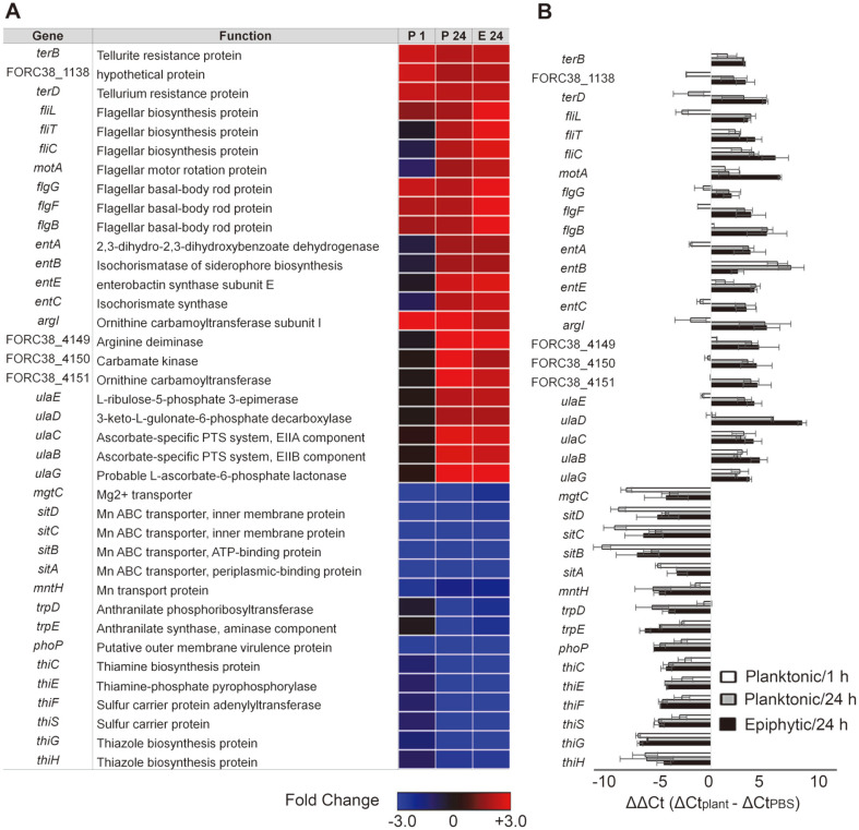
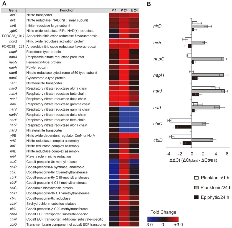
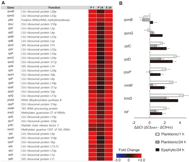
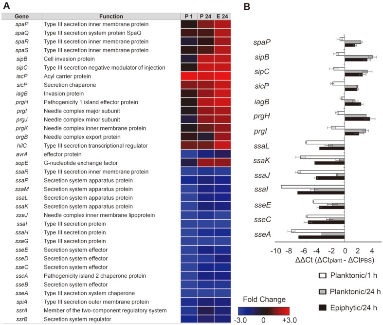
Salmonellosis is a form of gastroenteritis caused by Salmonella infection. The main transmission route of salmonellosis has been identified as poorly cooked meat and poultry products contaminated with Salmonella. However, in recent years, the number of outbreaks attributed to contaminated raw produce has increased dramatically. To understand how Salmonella adapts to produce, transcriptomic analysis was conducted on Salmonella enterica serovar Virchow exposed to fresh-cut radish greens. Considering the different Salmonella lifestyles in contact with fresh produce, such as motile and sessile lifestyles, total RNA was extracted from planktonic and epiphytic cells separately. Transcriptomic analysis of S. Virchow cells revealed different transcription profiles between lifestyles. During bacterial adaptation to fresh-cut radish greens, planktonic cells were likely to shift toward anaerobic metabolism, exploiting nitrate as an electron acceptor of anaerobic respiration, and utilizing cobalamin as a cofactor for coupled metabolic pathways. Meanwhile, Salmonella cells adhering to plant surfaces showed coordinated upregulation in genes associated with translation and ribosomal biogenesis, indicating dramatic cellular reprogramming in response to environmental changes. In accordance with the extensive translational response, epiphytic cells showed an increase in the transcription of genes that are important for bacterial motility, nucleotide transporter/metabolism, cell envelope biogenesis, and defense mechanisms. Intriguingly, Salmonella pathogenicity island (SPI)-1 and SPI-2 displayed up- and downregulation, respectively, regardless of lifestyles in contact with the radish greens, suggesting altered Salmonella virulence during adaptation to plant environments. This study provides molecular insights into Salmonella adaptation to plants as an alternative environmental reservoir.
Keywords: Salmonella enterica; epiphytic; planktonic; produce; transcriptomics.
Conflict of interest statement
The authors have no financial conflicts of interest to declare.
Figures

Similar articles
-
Understanding Comprehensive Transcriptional Response of Salmonella enterica spp. in Contact with Cabbage and Napa Cabbage.J Microbiol Biotechnol. 2018 Nov 28;28(11):1896-1907. doi: 10.4014/jmb.1806.06018. J Microbiol Biotechnol. 2018. PMID: 30270599
-
glnA Truncation in Salmonella enterica Results in a Small Colony Variant Phenotype, Attenuated Host Cell Entry, and Reduced Expression of Flagellin and SPI-1-Associated Effector Genes.Appl Environ Microbiol. 2018 Jan 2;84(2):e01838-17. doi: 10.1128/AEM.01838-17. Print 2018 Jan 15. Appl Environ Microbiol. 2018. PMID: 29150501 Free PMC article.
-
Survival and transcriptomic response of Salmonella enterica on fresh-cut fruits.Int J Food Microbiol. 2021 Jun 16;348:109201. doi: 10.1016/j.ijfoodmicro.2021.109201. Epub 2021 Apr 20. Int J Food Microbiol. 2021. PMID: 33930836
-
Salmonella-how a metabolic generalist adopts an intracellular lifestyle during infection.Front Cell Infect Microbiol. 2015 Jan 29;4:191. doi: 10.3389/fcimb.2014.00191. eCollection 2014. Front Cell Infect Microbiol. 2015. PMID: 25688337 Free PMC article. Review.
-
Salmonella pathogenicity island 1(SPI-1) at work.Curr Microbiol. 2013 Jun;66(6):582-7. doi: 10.1007/s00284-013-0307-8. Epub 2013 Jan 31. Curr Microbiol. 2013. PMID: 23370732 Review.
Cited by
-
Comparative Gene Expression Analysis of Salmonella Typhimurium DT104 in Ground Chicken Extract and Brain Heart Infusion Broth.Microorganisms. 2024 Jul 18;12(7):1461. doi: 10.3390/microorganisms12071461. Microorganisms. 2024. PMID: 39065229 Free PMC article.
-
Biological Function of Prophage-Related Gene Cluster ΔVpaChn25_RS25055~ΔVpaChn25_0714 of Vibrio parahaemolyticus CHN25.Int J Mol Sci. 2024 Jan 23;25(3):1393. doi: 10.3390/ijms25031393. Int J Mol Sci. 2024. PMID: 38338671 Free PMC article.
References
-
- Kroupitski Y, Golberg D, Belausov E, Pinto R, Swartzberg D, Granot D, et al. Internalization of Salmonella enterica in leaves is induced by light and involves chemotaxis and penetration through open stomata. Appl. Environ. Microbiol. 2009;75:6076–6086. doi: 10.1128/AEM.01084-09. - DOI - PMC - PubMed
MeSH terms
Substances
LinkOut - more resources
Full Text Sources
Medical
Miscellaneous