. 2020 Aug 24;11(1):366.
doi: 10.1186/s13287-020-01883-5.

COCO enhances the efficiency of photoreceptor precursor differentiation in early human embryonic stem cell-derived retinal organoids

Affiliations

COCO enhances the efficiency of photoreceptor precursor differentiation in early human embryonic stem cell-derived retinal organoids

Deng Pan et al. Stem Cell Res Ther. .

Abstract

Background: Significant progress has been made in cell replacement therapy for neural retinal diseases using retinal cells differentiated from human pluripotent stem cells. Low tumorigenicity and the ability to mature to form synaptic junctions make precursor cells a promising donor source. Here, we attempted to improve the yield of photoreceptor precursor cells in three-dimensional retinal organoids from human embryonic stem cells (hESCs).

Methods: A CRX-tdTomato-tagged hESC line was generated to track retinal precursors in 3D retinal organoids. COCO, a multifunctional antagonist of the Wnt, TGF-β, and BMP pathways, was employed to 3D organoid differentiation schemes for enhanced photoreceptor precursor cells. Organoid fluorescence intensity measurement was used to monitor retinalization tendency with the number of precursors further checked by flow cytometry. Signature gene expression during organoid differentiation were assessed by qPCR and immunocytochemistry after COCO supplementation.

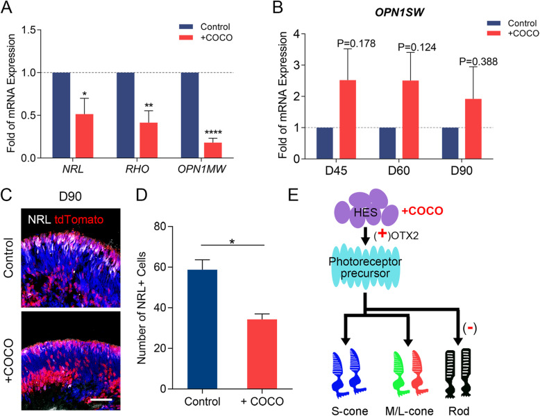
Results: CRX-positive cells can be spatiotemporally tracked by tdTomato without affecting retinalization during retinal organoid differentiation. Fluorescence intensity of organoids, which turned out highly consistent with flow cytometry measurement, allowed us to determine the differentiation efficiency of precursors during organoid culturing directly. Using COCO as an auxiliary supplement, rather than alone, can yield an increased number of photoreceptor precursors in the early stage of organoid differentiation. Over a longer time-frame, photoreceptor precursors enhanced their fate of cones and decreased fate of rods after treatment with COCO.

Conclusions: Tracing with the CRX-reporter system showed that in retinal organoids derived from human pluripotent stem cells, COCO increased the differentiation efficiency of photoreceptor precursors and cones.

Keywords: 3D; COCO; CRX; Cone; Fluorescent labeling; Photoreceptor precursor; Retinal organoid.


Conflict of interest statement

The authors declare no conflicts of interest.

Figures

Fig. 1
Fig. 1
Generation of reporter knock-in CRXp-tdTomato cell line. a Schematic diagram showing the targeting strategy of the insertion site. tdTomato cDNA sequence was fused in-frame into CRX behind start codon. b Overall 3D organoids fluorescent and bright field images on D45, D60, D90, and D120. A typical fluorescence intensity increasement along with differentiation time is presented. c Representative flow cytometry analysis in D45, D60, D90, and D120 organoids. Black and red represent the organoids derived from control and knock cell line, respectively. d Overall fluorescence intensity of organoids quantified by ImageJ, data are presented as the mean ± SEM (n = 27, 14, 11, and 10, respectively). e Proportion of tdTomato-positive cells in 3D organoids counted by flow cytometry, data are presented as the mean ± SEM (n = 6, 6, 8, and 3, respectively). f Comparison of increasement tendency between flow cytometry analysis and fluorescence intensity. g Relationship between fluorescence intensity and the number of tdTomato-positive cell in 3D organoids
Fig. 2
Fig. 2
tdTomato accurately traced photoreceptor precursor cells in 3D retinal organoids. a Immunocytochemistry with anti-CRX showing high correlation between tdTomato and CRX in D45, D60, and D90 organoids. Scale bar, 50 μm. b Quantification of anti-CRX-positive cells and tdTomato-positive cells at different stages of differentiation. c Immunocytochemical analysis between H9 and CRXp-tdTomato after retinal organoid differentiation indicates an undisturbed progenitor, ganglion cell, and rod development. Scale bar, 50 μm. d Quantification of ganglion, progenitor, and rod cells in D45, D60, and D90 organoids
Fig. 3
Fig. 3
Acceleration of photoreceptor precursor cells after COCO supplement. a Schematic illustration outlining the differentiation protocol to improve precursor cells with COCO. b Fluorescent organoids inducing efficiency supplied by COCO and COCO with IGF1 at different developmental stages. Scale bar, 100 μm. c Fluorescence intensity analysis in D45, D60, D90, and D120 organoids of COCO group (n = 27, 30, 17, 5, respectively) and COCO+IGF1 group (n = 27, 23, 10, 4, respectively) versus control retinal organoids (n = 17, 14, 11, 10, respectively). d Photoreceptor precursor cells between COCO group and control. e Immunohistochemistry with anti-RCVRN antibody on D45 and D60 organoids. Scale bar, 50 μm
Fig. 4
Fig. 4
Characterization of marker gene expression in 3D organoids after COCO supplement. a Percentage mRNA expression of PAX6, VSX2, and BRN3B transcripts in early-stage differentiation analyzed by qPCR in COCO supplement and control. b Immunostaining of gene products showed in a. Scale bar, 50 μm. c Percentage mRNA expression of photoreceptor related OTX2 and RCVRN transcripts. d Percentage mRNA expression of all subtypes of cones expressed ARR3 and M-cone expressed RXRG and THRB transcripts in COCO supplement and control. Data are expressed as mean ± SEM. e Immunocytochemical analysis with anti-OTX2 and anti-RXRG. Scale bar, 50 μm. f, g Statistic analysis of OTX2+ and OTX2+ cells in organoids. *P < 0.05, **P < 0.01, ns, not significant
Fig. 5
Fig. 5
Effects on photoreceptors at middle developmental stage after COCO supplement. a Percentage mRNA expression of NRL, RHO, and OPN1MW transcripts in D90 organoids analyzed by qPCR in COCO supplement and control. b Percentage mRNA expression of OPN1SW transcripts in D45, D60, and D90 organoids. c, d Immunocytochemical analysis and quantification for NRL. Data are expressed as mean ± SEM. Scale bar, 50 μm. e The proposed effect of COCO at early developmental stage intervention in 3D retinalization. *P < 0.05

References

    1. Jayakody SA, Gonzalez-Cordero A, Ali RR, Pearson RA. Cellular strategies for retinal repair by photoreceptor replacement. Prog Retin Eye Res. 2015;46:31–66. - PubMed
    1. Zhao C, Wang Q, Temple S. Stem cell therapies for retinal diseases: recapitulating development to replace degenerated cells. Development. 2017;144(8):1368–1381. - PMC - PubMed
    1. Jin ZB, Gao ML, Deng WL, Wu KC, Sugita S, Mandai M, et al. Stemming retinal regeneration with pluripotent stem cells. Prog Retin Eye Res. 2019;69:38–56. - PubMed
    1. Gagliardi G, Ben M'Barek K, Chaffiol A, Slembrouck-Brec A, Conart JB, Nanteau C, et al. Characterization and transplantation of CD73-positive photoreceptors isolated from human iPSC-derived retinal organoids. Stem Cell Rep. 2018;11(3):665–680. - PMC - PubMed
    1. MacLaren RE, Pearson RA, MacNeil A, Douglas RH, Salt TE, Akimoto M, et al. Retinal repair by transplantation of photoreceptor precursors. Nature. 2006;444(7116):203–207. - PubMed

Publication types