. 2020 Aug 7:2020:3840124.
doi: 10.1155/2020/3840124. eCollection 2020.

Prevention of Oxygen-Induced Inflammatory Lung Injury by Caffeine in Neonatal Rats

Affiliations

Prevention of Oxygen-Induced Inflammatory Lung Injury by Caffeine in Neonatal Rats

Stefanie Endesfelder et al. Oxid Med Cell Longev. .

Abstract

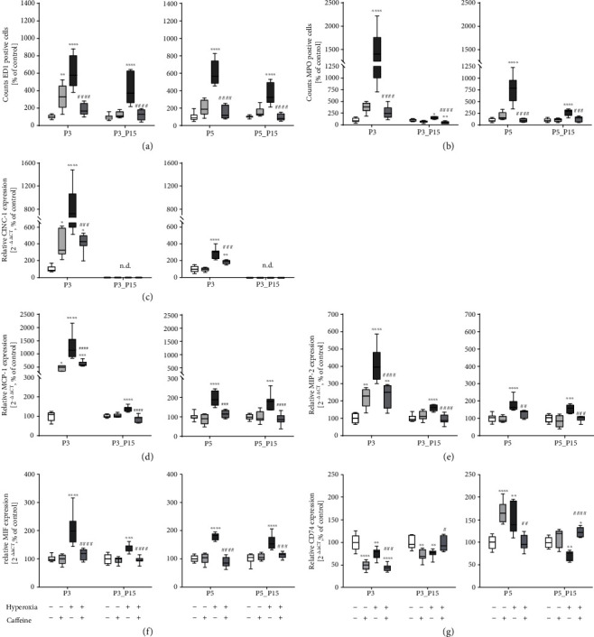
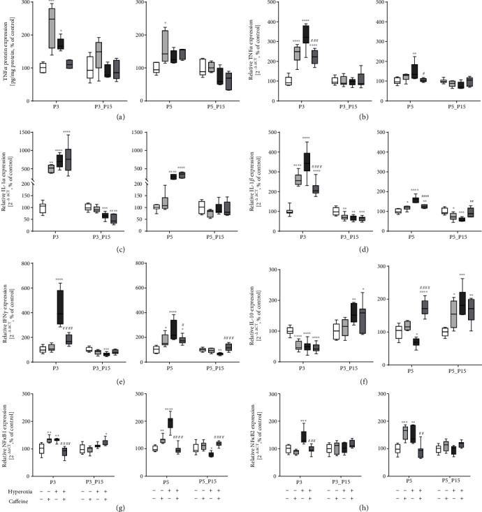
Background: Preterm birth implies an array of respiratory diseases including apnea of prematurity and bronchopulmonary dysplasia (BPD). Caffeine has been introduced to treat apneas but also appears to reduce rates of BPD. Oxygen is essential when treating preterm infants with respiratory problems but high oxygen exposure aggravates BPD. This experimental study is aimed at investigating the action of caffeine on inflammatory response and cell death in pulmonary tissue in a hyperoxia-based model of BPD in the newborn rat. Material/Methods. Lung injury was induced by hyperoxic exposure with 80% oxygen for three (P3) or five (P5) postnatal days with or without recovery in ambient air until postnatal day 15 (P15). Newborn Wistar rats were treated with PBS or caffeine (10 mg/kg) every two days beginning at the day of birth. The effects of caffeine on hyperoxic-induced pulmonary inflammatory response were examined at P3 and P5 immediately after oxygen exposure or after recovery in ambient air (P15) by immunohistological staining and analysis of lung homogenates by ELISA and qPCR.

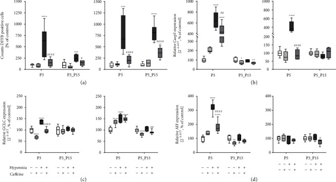
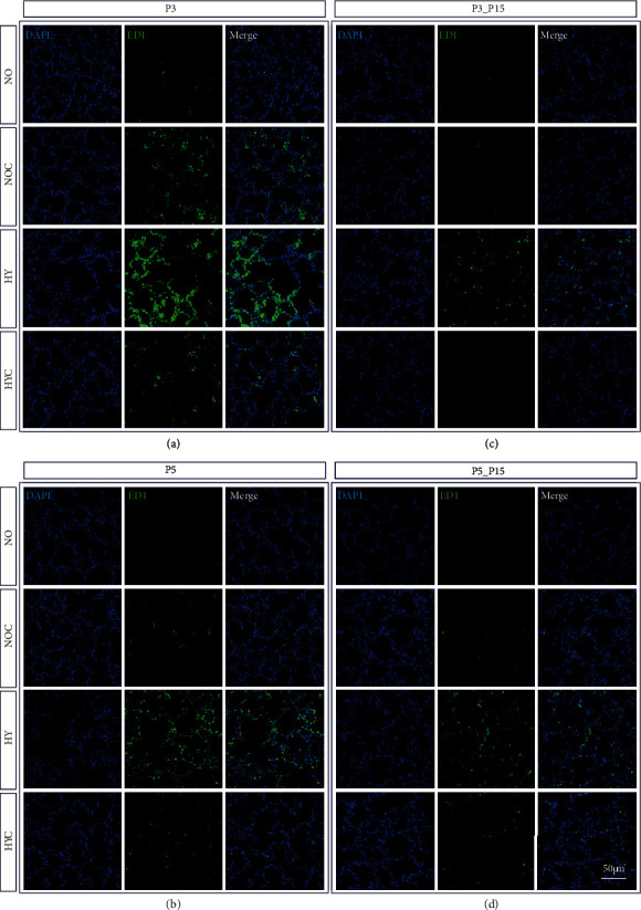
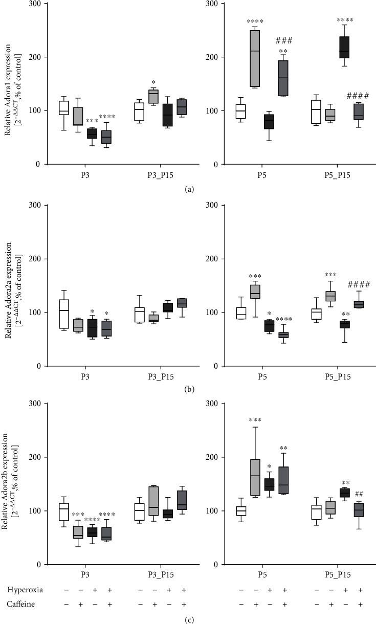
Results: Treatment with caffeine significantly attenuated changes in hyperoxia-induced cell death and apoptosis-associated factors. There was a significant decrease in proinflammatory mediators and redox-sensitive transcription factor NFκB in the hyperoxia-exposed lung tissue of the caffeine-treated group compared to the nontreated group. Moreover, treatment with caffeine under hyperoxia modulated the transcription of the adenosine receptor (Adora)1. Caffeine induced pulmonary chemokine and cytokine transcription followed by immune cell infiltration of alveolar macrophages as well as increased adenosine receptor (Adora1, 2a, and 2b) expression.

Conclusions: The present study investigating the impact of caffeine on the inflammatory response, pulmonary cell degeneration and modulation of adenosine receptor expression, provides further evidence that caffeine acts as an antioxidative and anti-inflammatory drug for experimental oxygen-mediated lung injury. Experimental studies may broaden the understanding of therapeutic use of caffeine in modulating detrimental mechanisms involved in BPD development.


Conflict of interest statement

The authors declare that there is no conflict of interest.

Figures

Figure 1
Figure 1
Representative photomicrographs of immunohistochemical staining of DFFB in the lungs of rat pups exposed to normoxia (NO) or hyperoxia (HY) compared to rat pups treated with caffeine (NOC, HYC). Examinations were performed at postnatal day 3 (P3 (a)) and P5 (b), or after recovery after 3-day exposure at P15 (c) or after 5-day exposure at P15 (d). Immunofluorescent images indicated DFFB (green) and nuclei (blue, DAPI). Scale bars represent 50 μm.
Figure 2
Figure 2
Quantitative analysis of (a) DFFB+ cell counts showed a marked increase in lung tissue samples of rat pups after acute oxygen exposure at P3 and P5 (deep dark grey bars) whereas caffeine treatment reduced apoptotic cells (dark grey bars). Cell death rate remained elevated even after recovery (P15). Caffeine treatment under room air (light grey bars) demonstrated no modulation of cell death. Hyperoxia within the first days of life accompanied by an increased cell death rate led to enhanced gene expression of (b) Casp3, (c) GCLC, and (d) AIF. Caffeine counteracted this. Quantification of lung homogenates was performed with qPCR for 3 days' postnatal oxygen exposure (P3) and recovery (P3_P15) and 5 days' postnatal oxygen exposure (P5) and recovery (P5_P15), respectively. Data are normalized to the level of rat pups exposed to normoxia at each time point (control 100%, white bars), and the 100% values are 4.3 (P3), 1.7 (P3_P15), 3.1 (P5), and 0.7 (P5_P15) cells per mm2, respectively. n = 6-8/group. p < 0.05, ∗∗p < 0.01, ∗∗∗p < 0.001, and ∗∗∗p < 0.0001 vs. control; #p < 0.05, ##p < 0.01, and ####p < 0.0001 vs. hyperoxia (ANOVA, Kruskal-Wallis, Dunn's post hoc test).
Figure 3
Figure 3
Representative micrographs of immunohistochemical staining of ED1 (CD68) in the lungs of rat pups exposed to normoxia (NO) or hyperoxia (HY) compared to rat pups treated with caffeine (NOC, HYC). Examinations were performed at postnatal day 3 (P3 (a)) and P5 (b), or after recovery after 3-day exposure at P15 (c) or after 5-day exposure at P15 (d). Immunofluorescent images indicated ED1 (green) and nuclei (blue, DAPI). Scale bars represent 100 μm.
Figure 4
Figure 4
Representative micrographs of immunohistochemical staining of MPO in the lungs of rat pups exposed to normoxia (NO) or hyperoxia (HY) compared to rat pups treated with caffeine (NOC, HYC). Examinations were performed at postnatal day 3 (P3 (a)) and P5 (b), or after recovery after 3-day exposure at P15 (c) or after 5-day exposure at P15 (d). Immunofluorescent images indicated MPO (green) and nuclei (blue, DAPI). Scale bars represent 100 μm.
Figure 5
Figure 5
Quantitative analysis of (a) ED1+ and (b) MPO+ cell counts showed a marked accumulation in lung tissue samples of rat pups after acute oxygen exposure at P3 and P5 (deep dark grey bars) whereas caffeine treatment inhibited (a) macrophage (ED1) and (b) neutrophil (MPO) infiltration (dark grey bars). Macrophage infiltration remained elevated even after recovery (P15). Caffeine treatment under room air (light grey bars) increased counts of macrophages at P3. Hyperoxia exposure of newborn rat pups, characterized by an overwhelming immune cell influx, led to increased chemokine gene expression of (c) CINC-1, (d) MCP-1, and (e) MIP-2. Chemokine transcription is inhibited by caffeine. Caffeine under normoxic exposure increased chemokine transcription at P3. CINC-1 is not detectable (n.d.) at P15. Caffeine alleviated the hyperoxia-induced gene expression of (f) MIF, whereas hyperoxia modulated expression of the MIF receptor (g) CD74, and caffeine counteracted this. Quantification of lung homogenates was performed with qPCR for 3 days' postnatal oxygen exposure (P3) and recovery (P3_P15) and 5 days' postnatal oxygen exposure (P5) and recovery (P5_P15), respectively. Data are normalized to the level of rat pups exposed to normoxia at each time point (control 100%, white bars), and the 100% values for ED1/MPO are 22.4/12.7 (P3), 7.6/12.5 (P3_P15), 15.2/12.0 (P5), and 7.1/10.9 (P5_P15) cells per mm−2, respectively. n = 6-8/group. p < 0.05, ∗∗p < 0.01, ∗∗∗p < 0.001, and ∗∗∗∗p < 0.0001 vs. control; #p < 0.05, ##p < 0.01, ###p < 0.001, and ####p < 0.0001 vs. hyperoxia (ANOVA, Kruskal-Wallis, Dunn's post hoc test).
Figure 6
Figure 6
Hyperoxia exposure of newborn rat pups is characterized by modulation of cytokine response and regulation of redox-sensitive transcription factors. High oxygen led to increased proinflammatory cytokine expression of (a) TNFα (protein), (b) TNFα (RNA), (c) IL-1α, (d) IL-1β, and (e) IFNγ. Anti-inflammatory cytokine expression of (f) IL-10 is suppressed and redox-sensitive transcription factors (g) NFκB1 and (h) NFκB2 are increased. Caffeine counteracted this. Caffeine under normoxic exposure demonstrated a massive modulation of proinflammatory cytokine expression. Quantification of lung homogenates by qPCR for 3 days' postnatal oxygen exposure (P3) and recovery (P3_P15) and 5 days' postnatal oxygen exposure (P5) and recovery (P5_P15), respectively. Data are normalized to the level of rat pups exposed to normoxia at each time point (control 100%, white bars). The 100% values of TNFα protein are 4.4 (P3), 5.6 (P3_P15), 3.5 (P5), and 7.2 (P5_P15) pg per ml, respectively. n = 7-8/group (qPCR); n = 5/group (ELISA). p < 0.05, ∗∗p < 0.01, ∗∗∗p < 0.001, and ∗∗∗∗p < 0.0001 vs. control; #p < 0.05, ##p < 0.01, and ####p < 0.0001 vs. hyperoxia (ANOVA, Kruskal-Wallis, Dunn's post hoc testq).
Figure 7
Figure 7
Hyperoxia and caffeine modulated gene expression of adenosine receptor subtypes (a) Adora1, (b) Adora2a, and (c) Adora2b. Quantification of lung homogenates was performed with qPCR for 3 days' postnatal oxygen exposure (P3) and recovery (P3_P15) and 5 days' postnatal oxygen exposure (P5) and recovery (P5_P15), respectively. Data are normalized to the level of rat pups exposed to normoxia at each time point (control 100%, white bars). n = 7-8/group. p < 0.05, ∗∗p < 0.01, ∗∗∗p < 0.001, and ∗∗∗∗p < 0.0001 vs. control; ##p < 0.01, ###p < 0.001, and ####p < 0.0001 vs. hyperoxia (ANOVA, Kruskal-Wallis, Dunn's post hoc test).

References

    1. Panfoli I., Candiano G., Malova M., et al. Oxidative stress as a primary risk factor for brain damage in preterm newborns. Frontiers in Pediatrics. 2018;6:p. 369. doi: 10.3389/fped.2018.00369. - DOI - PMC - PubMed
    1. Castillo A., Sola A., Baquero H., et al. Pulse oxygen saturation levels and arterial oxygen tension values in newborns receiving oxygen therapy in the neonatal intensive care unit: is 85% to 93% an acceptable range? Pediatrics. 2008;121(5):882–889. doi: 10.1542/peds.2007-0117. - DOI - PubMed
    1. Negi R., Pande D., Kumar A., Khanna R. S., Khanna H. D. Evaluation of biomarkers of oxidative stress and antioxidant capacity in the cord blood of preterm low birth weight neonates. The Journal of Maternal-Fetal & Neonatal Medicine. 2012;25(8):1338–1341. doi: 10.3109/14767058.2011.633672. - DOI - PubMed
    1. Perrone S., Bracciali C., di Virgilio N., Buonocore G. Oxygen use in neonatal care: a two-edged sword. Frontiers in Pediatrics. 2017;4:143–143. doi: 10.3389/fped.2016.00143. - DOI - PMC - PubMed
    1. Parkinson J. R. C., Hyde M. J., Gale C., Santhakumaran S., Modi N. Preterm birth and the metabolic syndrome in adult life: a systematic review and meta-analysis. Pediatrics. 2013;131(4):e1240–e1263. doi: 10.1542/peds.2012-2177. - DOI - PubMed