. 2020 Aug 1:2020:4074832.
doi: 10.1155/2020/4074832. eCollection 2020.

Short-Chain Fatty Acids Ameliorate Diabetic Nephropathy via GPR43-Mediated Inhibition of Oxidative Stress and NF- κ B Signaling

Affiliations

Short-Chain Fatty Acids Ameliorate Diabetic Nephropathy via GPR43-Mediated Inhibition of Oxidative Stress and NF- κ B Signaling

Wei Huang et al. Oxid Med Cell Longev. .

Abstract

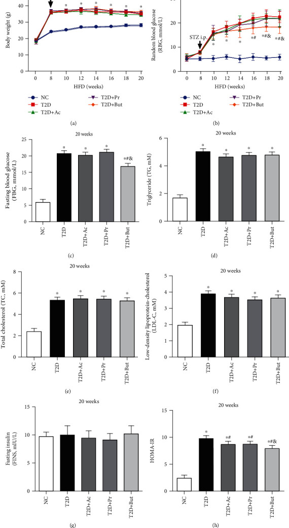
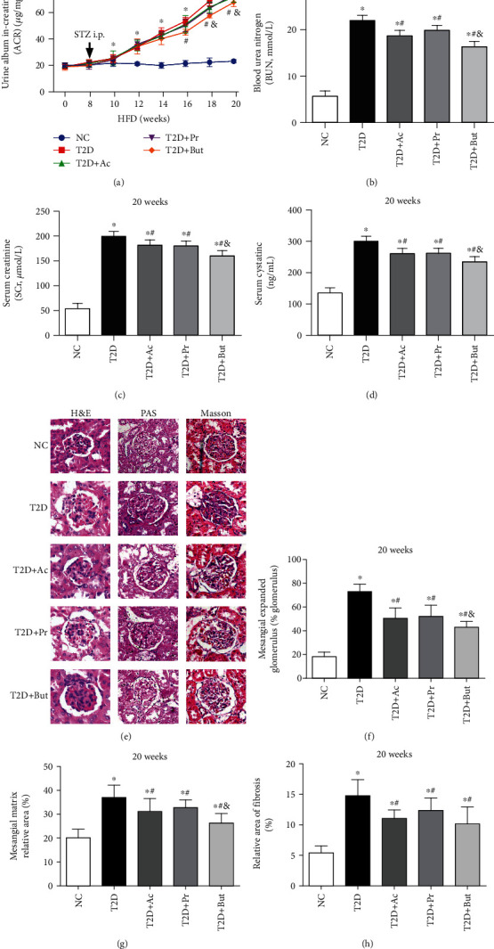
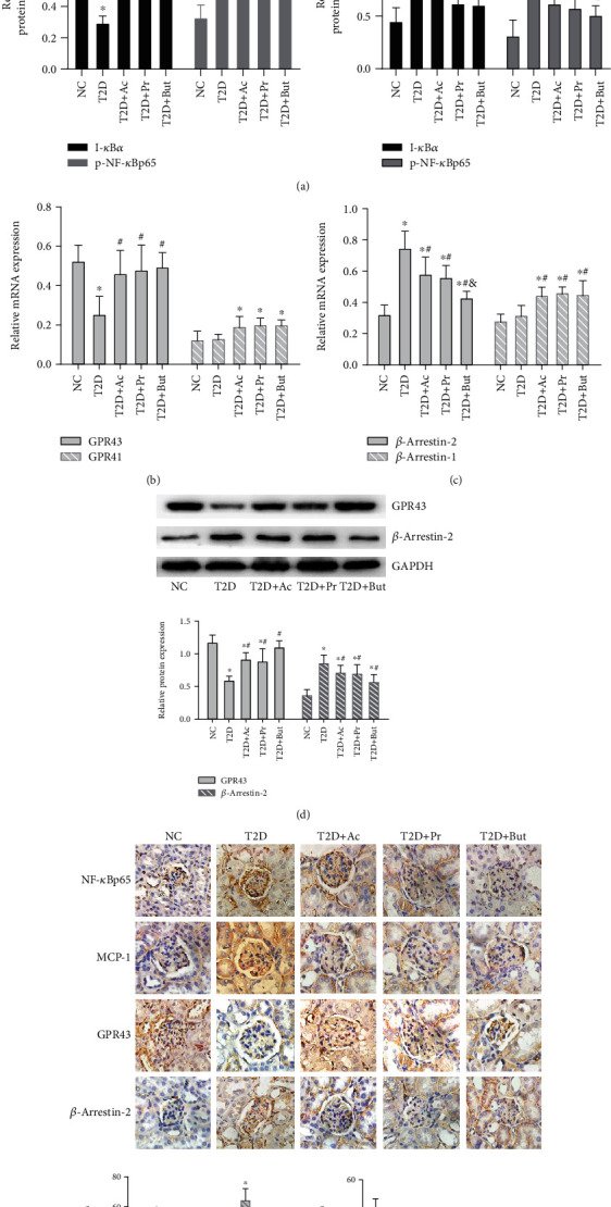
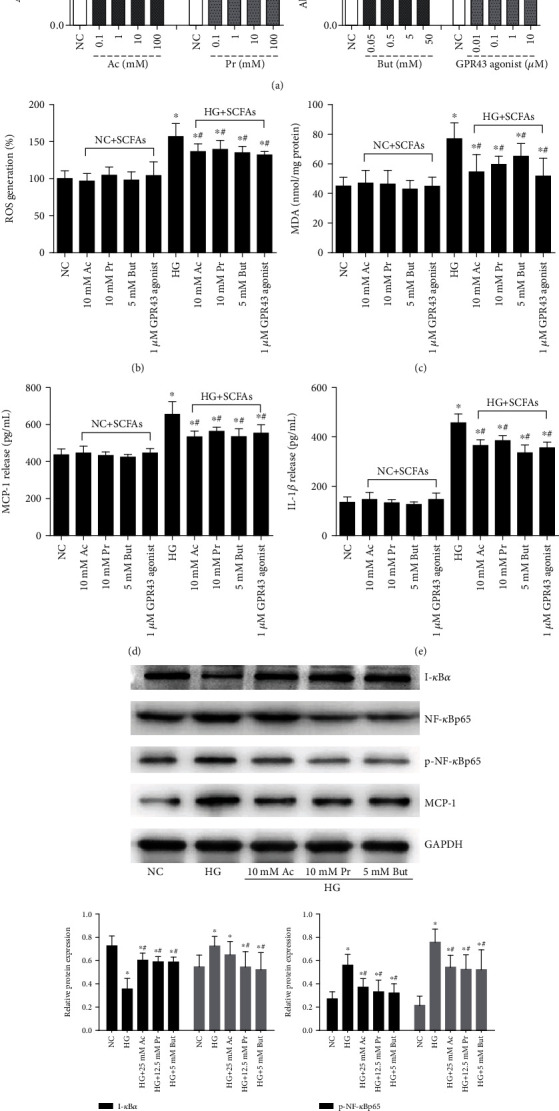
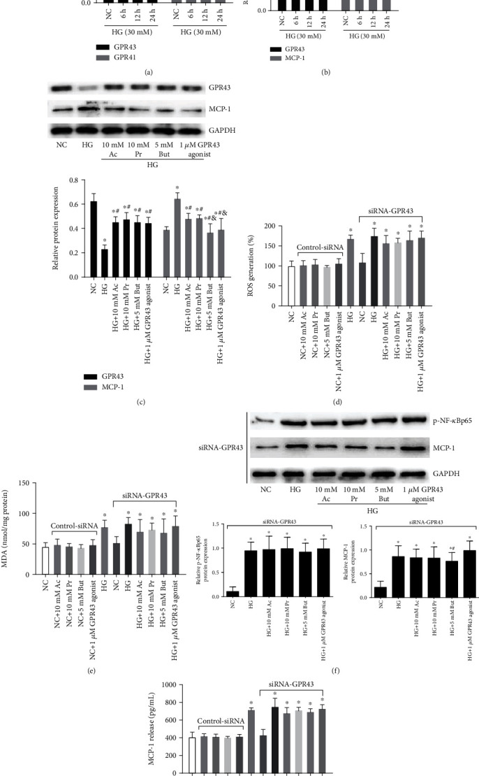
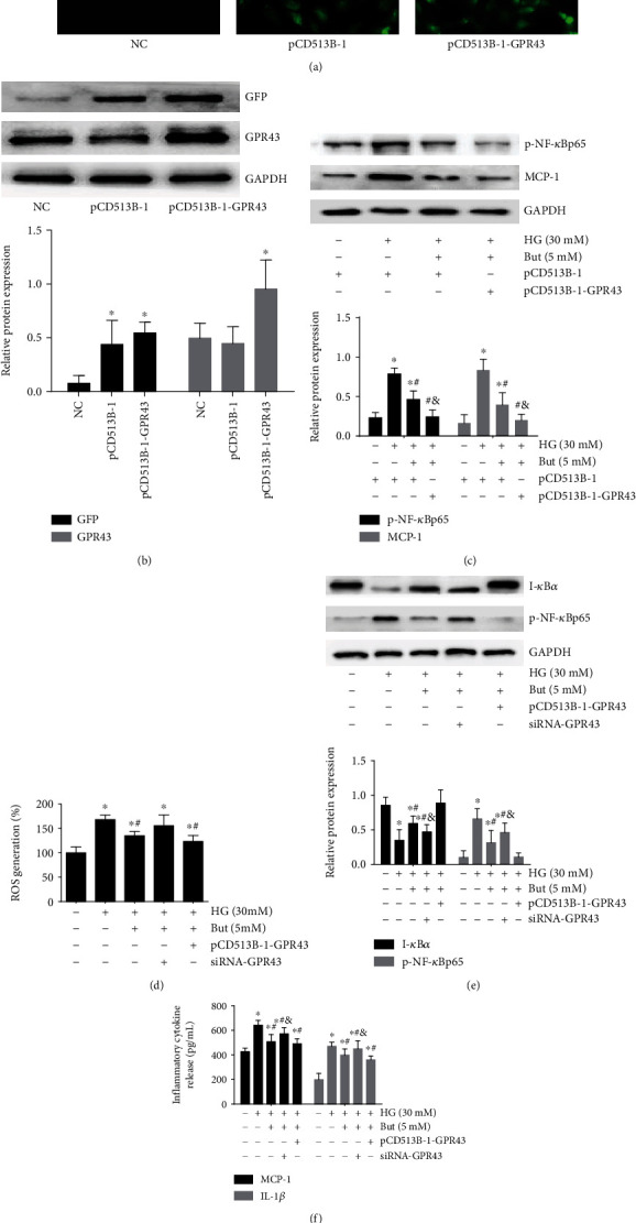
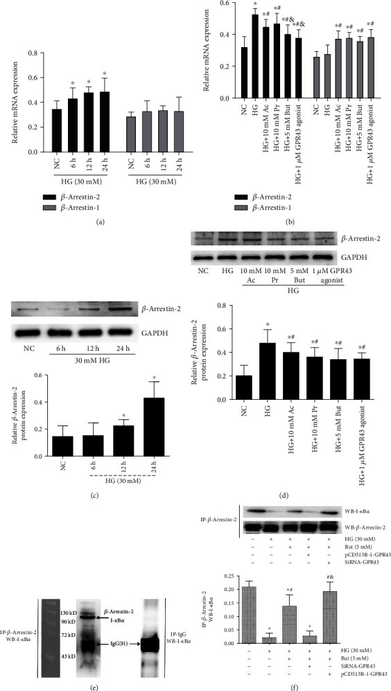
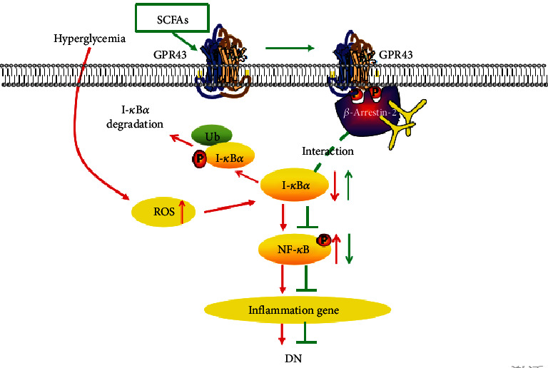
Diabetic nephropathy (DN) is a chronic low-grade inflammatory disease. Oxidative stress and nuclear factor kappa B (NF-κB) signaling play an important role in the pathogenesis of DN. Short-chain fatty acids (SCFAs) produced from carbohydrate fermentation in the gastrointestinal tract exert positive regulatory effects on inflammation and kidney injuries. However, it is unclear whether SCFAs can prevent and ameliorate DN. In the present study, we evaluated the role and mechanism of the three main SCFAs (acetate, propionate, and butyrate) in high-fat diet (HFD) and streptozotocin- (STZ-) induced type2 diabetes (T2D) and DN mouse models and in high glucose-induced mouse glomerular mesangial cells (GMCs), to explore novel therapeutic strategies and molecular targets for DN. We found that exogenous SCFAs, especially butyrate, improved hyperglycemia and insulin resistance; prevented the formation of proteinuria and an increase in serum creatinine, urea nitrogen, and cystatin C; inhibited mesangial matrix accumulation and renal fibrosis; and blocked NF-κB activation in mice. SCFAs also inhibited high glucose-induced oxidative stress and NF-κB activation and enhanced the interaction between β-arrestin-2 and I-κBα in GMCs. Specifically, the beneficial effects of SCFAs were significantly facilitated by the overexpression GPR43 or imitated by a GPR43 agonist but were inhibited by siRNA-GPR43 in GMCs. These results support the conclusion that SCFAs, especially butyrate, partially improve T2D-induced kidney injury via GPR43-mediated inhibition of oxidative stress and NF-κB signaling, suggesting SCFAs may be potential therapeutic agents in the prevention and treatment of DN.


Conflict of interest statement

The authors declare that they have no conflict of interest.

Figures

Figure 1
Figure 1
SCFAs ameliorated hyperglycemia and insulin resistance of experimental T2D. Mice were subjected to a high-fat diet (HFD) for 8 weeks, intraperitoneally (i.p.) injected with STZ, and then treated with three main SCFAs, acetate (Ac), propionate (Pr), and butyrate (But), for 12 weeks. Body weight (BW) (a) and random blood glucose (RBG) (b) were measured every 2 weeks; fasting blood glucose (FBG) (c), total cholesterol (TC) (d), total glyceride (TG) (e), low-density lipoprotein-cholesterol (LDL-C) (f), fasting insulin (FINS) (g), and homeostatic model assessment of insulin resistance (HOMA-IR) (h) values were measured at the 20th week of the experiment before sacrifice. p < 0.05 compared with the NC group; #p < 0.05 compared with the T2D group; &p < 0.05 compared with the Ac or Pr group.
Figure 2
Figure 2
SCFAs prevented the renal dysfunction and kidney injury. Urine ACR (a) of T2D mice were measured every 2 weeks, and blood urea nitrogen (BUN) (b), serum creatinine (SCr) (c), and serum cystatin C (d) were assayed at the 20th week of the experiment. Histopathological examination of renal tissues was by H&E, PAS, and Masson's trichrome staining (400x) (e). Mesangial expansion (f), glomerular tuft (g), and the accumulation of collagen (h) were measured. Ac: acetate; Pr: propionate; But: butyrate; p < 0.05 compared with the NC group; #p < 0.05 compared with the T2D group; &p < 0.05 compared with the Ac or Pr group.
Figure 3
Figure 3
SCFAs inhibited T2D-induced NF-κB activation and regulate GPR43-β-arrestin-2 signaling. (a) Western blotting revealed the expression of I-κBα, p-NF-κBp65, MCP-1, and IL-1β in T2D kidney tissue after SCFA treatment. (b, c) qRT-PCR of GPR43, GPR41, β-arrestin-2, and β-arrestin-1 in kidney tissue after SCFA treatment. (d) Western blotting-based assays for the expression of GPR43 and β-arrestin-2 in kidney tissue after SCFA treatment. (e) Immunohistochemistry- (400x) based assays for the expression of p-NF-κBp65, MCP-1, GPR43, and β-arrestin-2 after SCFA treatment. Ac: acetate; Pr: propionate; But: butyrate; p < 0.05 compared with the NC group; #p < 0.05 compared with the T2D group; &p < 0.05 compared with the Ac or Pr group.
Figure 4
Figure 4
SCFA treatment partially inhibited oxidative stress and NF-κB activation in high glucose-induced GMCs. (a) The effects of a concentration range of SCFAs or GPR43 agonist on GMC proliferation were analyzed by MTT assay. GMCs were stimulated with 30 mM high glucose in the presence of the indicated concentration of SCFAs or GPR43 agonist for 24 h. ROS (b), MDA (c), MCP-1 (d), and IL-1β (e) in the cell culture supernatant were evaluated by kit. The protein expression of I-κBα, NF-κBp65, p-NF-κBp65, and MCP-1 was assayed by western blotting (f). Ac: acetate group; Pr: propionate group; But: butyrate group; p < 0.05 compared with the NC group; #p < 0.05 compared with the HG group.
Figure 5
Figure 5
SCFA-mediated antioxidant and anti-inflammatory effects were partly reversed by siRNA-GPR43. qRT-PCR was performed to detect GPR43 and GPR41 mRNA levels. (b) Western blotting-based assay for the expression of GPR43 and MCP-1 after a 30 mM high glucose challenge for 6, 12, and 24 h. (c) The effects of indicated concentrations of SCFAs or a GPR43 agonist on GPR43 and MCP-1 expression were analyzed by western blotting. Following 30 mM high glucose for 24 h, SCFAs or a GPR43 agonist-ameliorated ROS (d), MDA (e), NF-κB signal (g), and MCP-1 (f) were significantly reversed by siRNA-GPR43. Ac: acetate; Pr: propionate; But: butyrate; p < 0.05 compared with the NC group; #p < 0.05 compared with the HG group. &p < 0.05 compared with the Ac or Pr group.
Figure 6
Figure 6
SCFA-mediated antioxidant and anti-inflammatory effects were significantly facilitated by GPR43 overexpression. Plasmids expressing GPR43 with pCD513B-1 (an N-terminal GFP tag) and pCD513B-1 control plasmid that expresses GFP but cannot overexpress GPR43 were constructed to determine the effect of overexpressed GPR43 and GFP protein in GMCs according to fluorescence images (200x) (a) and western blotting (b). Following 30 mM high glucose for 24 h, 5 mM But-mediated inhibition of p-NF-κBp65 and MCP-1 protein expression (c) was significantly facilitated by GPR43 overexpression. The But-mediated inhibition of ROS (d) and p-NF-κBp65 (e) and MCP-1 and IL-1β release (f) were reversed by siRNA-GPR43 but facilitated by GPR43 overexpression. But butyrate: p < 0.05 compared with the NC group, #p < 0.05 compared with the HG group, and &p < 0.05 compared with the But group.
Figure 7
Figure 7
Interaction between β-arrestin-2 and I-κBα was induced by SCFAs via GPR43. (a) GMCs were treated with 30 mM high glucose for 6, 12, and 24 h. RT-PCR was performed to detect β-arrestin-2 and β-arrestin-1 mRNA levels. (b) The effects of indicated concentrations of SCFAs or an GPR43 agonist on β-arrestin-2 and β-arrestin-1 expression were analyzed by RT-PCR. (c) Western blot assay for the expression of β-arrestin-2 after 30 mM high glucose challenge for 6, 12, and 24 h. (d) High glucose-induced β-arrestin-2 expression was significantly reversed by SCFAs or GPR43 agonist. (e) The interaction between β-arrestin-2 and I-κBα under physiological conditions was detected by immunoprecipitation (IP) with anti-β-arrestin-2 antibody or normal mouse IgG antibody (negative control), followed by western blotting with an anti-I-κBα antibody. β-Arrestin-2 was conjugated with I-κBα in vitro. (f) The interaction between β-arrestin-2 and I-κBα was decreased by 30 mM high glucose but was reversed by 5 mM butyrate. And these butyrate-mediated effects were significantly reversed by siRNA-GPR43 but were facilitated by overexpressed GPR43. IgG-H marks the IgG heavy chain. But butyrate: p < 0.05 compared with the NC group, #p < 0.05 compared with the HG group, and &p < 0.05 compared with the HG+But group. Ac: acetate; Pr: propionate.
Figure 8
Figure 8
Overview on the effects of SCFAs on oxidative stress and NF-κB activation in DN. High glucose induces the production of ROS and the polyubiquitination of phosphorylated I-κBα, followed by NF-κB activation and the expression of various inflammatory cytokines that are important factors in the development of DN (red arrows). However, SCFAs inhibit the oxidative stress and NF-κB inflammatory signaling possibly via activating GPR43 and increasing the interaction between β-arrestin-2 and I-κBα (green arrows), suggesting that SCFA-mediated GPR43-β-arrestin-2 signaling may be a novel and promising target for DN.

References

    1. Marshall C. B. Rethinking glomerular basement membrane thickening in diabetic nephropathy: adaptive or pathogenic? American Journal of Physiology. Renal Physiology. 2016;311(5):F831–F843. doi: 10.1152/ajprenal.00313.2016. - DOI - PMC - PubMed
    1. Kawanami D., Matoba K., Utsunomiya K. Signaling pathways in diabetic nephropathy. Histology and Histopathology. 2016;31(10):1059–1067. doi: 10.14670/HH-11-777. - DOI - PubMed
    1. Johnson S. A., Spurney R. F. Twenty years after ACEIs and ARBs: emerging treatment strategies for diabetic nephropathy. American Journal of Physiology. Renal Physiology. 2015;309(10):F807–F820. doi: 10.1152/ajprenal.00266.2015. - DOI - PMC - PubMed
    1. He M., Shi B. Gut microbiota as a potential target of metabolic syndrome: the role of probiotics and prebiotics. Cell & Bioscience. 2017;7(1) doi: 10.1186/s13578-017-0183-1. - DOI - PMC - PubMed
    1. Schönfeld P., Wojtczak L. Short- and medium-chain fatty acids in energy metabolism: the cellular perspective. Journal of Lipid Research. 2016;57(6):943–954. doi: 10.1194/jlr.R067629. - DOI - PMC - PubMed