

Endobronchial metastases from a primary embryonal carcinoma
- PMID: 32832088
- PMCID: PMC7438807
- DOI: 10.1002/rcr2.644
Endobronchial metastases from a primary embryonal carcinoma
Abstract
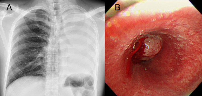
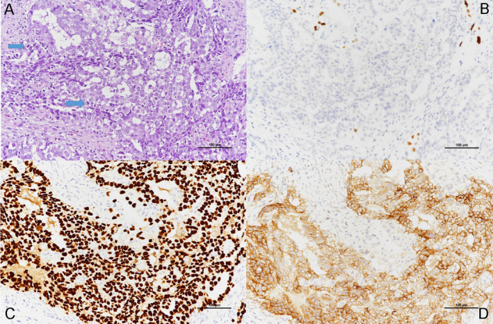
We report the case of a 24-year-old man who presented with chief complaints of shortness of breath and haemoptysis; chest radiography revealed complete collapse of the left lung. Bronchoscopy revealed an endobronchial tumour with complete obstruction of the left main bronchus. Cryosurgical excision was performed; tissue pathology confirmed the diagnosis of metastatic embryonal carcinoma. The patient underwent a right orchiectomy followed by a bleomycin + etoposide + cisplatin (BEP) chemotherapy regimen.
Keywords: Cryosurgery; embryonal carcinoma; endobronchial metastases; endobronchial tumour.
© 2020 The Authors. Respirology Case Reports published by John Wiley & Sons Australia, Ltd on behalf of The Asian Pacific Society of Respirology.
Figures
Similar articles
-
[Endobronchial metastasis of testicular embryonal cell carcinoma--a case report].Gan No Rinsho. 1983 Sep;29(11):1340-3. Gan No Rinsho. 1983. PMID: 6632215 Japanese.
-
[A case of testicular cancer in which complete response was achieved with chemotherapy including cisplatin, etoposide and bleomycin, and salvage surgery].Gan To Kagaku Ryoho. 1994 Mar;21(4):543-6. Gan To Kagaku Ryoho. 1994. PMID: 7510465 Japanese.
-
Urethral metastasis from primary embryonal carcinoma of testis - The first case report.Urol Case Rep. 2021 Jan 20;36:101572. doi: 10.1016/j.eucr.2021.101572. eCollection 2021 May. Urol Case Rep. 2021. PMID: 33532243 Free PMC article.
-
[Intra-arterial infusion chemotherapy for liver metastases of testicular tumors: report of two cases].Hinyokika Kiyo. 2000 Nov;46(11):823-7. Hinyokika Kiyo. 2000. PMID: 11193306 Review. Japanese.
-
[Bilateral and synchronous testicular teratoma: a case report and literature review].Cir Cir. 2015 Nov-Dec;83(6):527-31. doi: 10.1016/j.circir.2015.05.035. Epub 2015 Jul 9. Cir Cir. 2015. PMID: 26164135 Review. Spanish.
Cited by
-
Mimicking Metastasis: Endobronchial Tuberculosis Presenting as a Mass in a Pediatric Patient With Yolk Sac Tumor.Cureus. 2024 Jul 24;16(7):e65277. doi: 10.7759/cureus.65277. eCollection 2024 Jul. Cureus. 2024. PMID: 39184706 Free PMC article.
References
-
- Öztürk A, Aktaş Z, and Yılmaz A. 2016. Endobronchial metastasis of mixed germ cell tumors: two cases. Tuberk. Toraks 64(2):175–178. - PubMed
-
- Ikemura K, Lin DM, Martyn CP, et al. 2017. Endobronchial metastasis from extrapulmonary neoplasms: analysis of clinicopathologic features and cytological evaluation by bronchial brushing. Lung 195(5):595–599. - PubMed
-
- Moreira‐Meyer A, Bautista‐Herrera D, Hernández‐González M, et al. 2017. Endobronchial embryonal carcinoma. J. Bronchology Interv. Pulmonol. 24(2):148–152. - PubMed
-
- Özsu S, Erol MM, Oztuna F, et al. 2008. Endobronchial metastasis from testicular seminoma. Med. Princ. Pract. 17:493–495. - PubMed
Publication types
LinkOut - more resources
Full Text Sources
