Comparison between Conventional Decalcification and a Microwave-Assisted Method in Bone Tissue Affected with Mycetoma
- PMID: 32832156
- PMCID: PMC7422918
- DOI: 10.1155/2020/6561980
Comparison between Conventional Decalcification and a Microwave-Assisted Method in Bone Tissue Affected with Mycetoma
Abstract
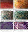
Mycetoma is a lifelong granulomatous disease of subcutaneous tissues and bones. Histopathology is a substantiated indicative method based on the assumption of a definitive diagnosis of mycetoma. It requires efficient processing of tissues including bone decalcification. The decalcification process must ensure complete removal of calcium and also a proper preservation of tissue and microorganisms' staining ability. Objectives. To compare the conventional method used in decalcification with the microwave method using different decalcification solutions. Different characteristics were tested, including the speed of decalcification and morphological and fungal preservation in bone tissue affected with mycetoma. Materials and Methods. Three decalcification solutions were employed to remove calcium from 50 bone tissue samples affected with mycetoma, including 10% neutral buffered EDTA (pH 7.4), 5% nitric acid, and 5% hydrochloric acid. Conventional and microwave methods were used. Haematoxylin-eosin (HE) stain, Gridley's stain, and Grocott hexamine-silver stain were employed to evaluate the bone and fungi morphologies. Results. The decalcification time of the conventional method compared with the microwave method with 10% EDTA (pH 7.4) took 120 hours and 29 hours, while 5% hydrochloric acid and 5% nitric acid took 8 hours and 3 hours, separately. Also, 10% EDTA is the best decalcifying agent for HE staining and fungal stains. 5% hydrochloric acid and 5% nitric acid can be used for fungal staining. Conclusion. The current study investigated the effects of different decalcifying agents as well as two decalcification procedures on the preservation of the bone structure and fungal staining, which will help to develop suitable protocols for the analyses of the bone tissue affected with mycetoma infection.
Copyright © 2020 Magdi Mansour Salih.
Conflict of interest statement
The author declares that there are no conflicts of interest.
Figures
Similar articles
-
Evaluation of Decalcification Techniques for Rat Femurs Using HE and Immunohistochemical Staining.Biomed Res Int. 2017;2017:9050754. doi: 10.1155/2017/9050754. Epub 2017 Jan 26. Biomed Res Int. 2017. PMID: 28246608 Free PMC article.
-
Comparison of routine decalcification methods with microwave decalcification of bone and teeth.J Oral Maxillofac Pathol. 2013 Sep;17(3):386-91. doi: 10.4103/0973-029X.125204. J Oral Maxillofac Pathol. 2013. PMID: 24574657 Free PMC article.
-
Comparison of Microwave Versus Conventional Decalcification of Teeth Using Three Different Decalcifying Solutions.J Lab Physicians. 2016 Jul-Dec;8(2):106-11. doi: 10.4103/0974-2727.180791. J Lab Physicians. 2016. PMID: 27365920 Free PMC article.
-
Microwave-Assisted Tissue Preparation for Rapid Fixation, Decalcification, Antigen Retrieval, Cryosectioning, and Immunostaining.Int J Cell Biol. 2016;2016:7076910. doi: 10.1155/2016/7076910. Epub 2016 Oct 20. Int J Cell Biol. 2016. PMID: 27840640 Free PMC article. Review.
-
Resolving the bone - optimizing decalcification in spatial transcriptomics and molecular pathology.J Histotechnol. 2025 Mar;48(1):68-77. doi: 10.1080/01478885.2024.2446038. Epub 2024 Dec 26. J Histotechnol. 2025. PMID: 39723974 Review.
Cited by
-
Protocol for assessing the structural architecture, integrity, and cellular composition of murine bone ex vivo.STAR Protoc. 2025 Jun 20;6(2):103843. doi: 10.1016/j.xpro.2025.103843. Epub 2025 May 24. STAR Protoc. 2025. PMID: 40413750 Free PMC article.
-
Arduino Automated Microwave Oven for Tissue Decalcification.Bioengineering (Basel). 2023 Jan 6;10(1):79. doi: 10.3390/bioengineering10010079. Bioengineering (Basel). 2023. PMID: 36671651 Free PMC article.
References
LinkOut - more resources
Full Text Sources


