Case Reports
. 2020 Aug 2:2020:8886936.
doi: 10.1155/2020/8886936. eCollection 2020.

Management of an Infected Vesicourachal Diverticulum in a 42-Year-Old Woman

Affiliations
Case Reports

Management of an Infected Vesicourachal Diverticulum in a 42-Year-Old Woman

Maria Erodotou et al. Case Rep Urol. .

Abstract

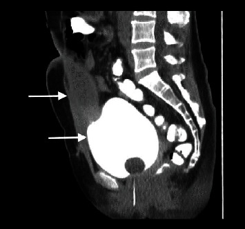
Urachal remnant anomalies are uncommon in adults and can be confused with a variety of clinical conditions when symptomatic or infected. Vesicourachal diverticulum is the rarest type, accounting for approximately 3% to 5% of congenital urachal anomalies. We report the case of a 42-year-old female patient, who presented to the emergency department with lower abdominal pain and a palpable abdominal mass. An infected vesicourachal diverticulum was diagnosed after imaging studies and was initially treated with intravenous antibiotic therapy and drainage of the urachal diverticulum to the urinary bladder through a JJ stent. Finally, the patient underwent open surgical excision of the urachal remnant. The postoperative course was uneventful, and the histopathological examination confirmed the diagnosis of vesicourachal diverticulum. We recommend drainage of an infected vesicourachal diverticulum through the bladder by JJ stent placement inside its lumen during cystoscopy, as an alternative to percutaneous drainage reported in the literature.


Conflict of interest statement

The authors declare that they have no conflict of interest.

Figures

Figure 1
Figure 1
Contrast–enhanced computed tomographic image showing a cystic lesion with peripheral enhancement (arrow), thus excluding the initial diagnosis of an incarcerated hernia.
Figure 2
Figure 2
Contrast–enhanced computed tomography (sagittal plane) showing the cystic lesion (arrow-above) arising from the bladder dome (arrow–below) and extending to the umbilical region, leading to a suspected diagnosis of an infected vesicourachal diverticulum.
Figure 3
Figure 3
Cystography showing the bladder filled with contrast (arrow-below) without diffusion of the contrast inside the vesicourachal diverticulum (arrow-above), probably due to infection and a small opening of its lumen.
Figure 4
Figure 4
Intraoperative image during the dissection of the urachal remnant (arrow).
Figure 5
Figure 5
Intraoperative image showing the vesicourachal diverticulum (arrow-above) arising from the anterosuperior margin of the bladder (arrow-below).
Figure 6
Figure 6
Surgical specimen of the resected vesicourachal diverticulum with a bladder cuff and the JJ stent in place (arrow).

References

    1. Parada Villavicencio C., Adam S. Z., Nikolaidis P., Yaghmai V., Miller F. H. Imaging of the urachus: anomalies, complications, and mimics. Radiographics. 2016;36(7):2049–2063. doi: 10.1148/rg.2016160062. - DOI - PubMed
    1. Passoni S., Guerra A., Marengo M. Laparoscopic treatment of an infected urachalcyst and diverticulum in a young adult: presentation of a case and review of the literature. International Journal of Surgery Case Reports. 2018;49:87–90. doi: 10.1016/j.ijscr.2018.06.018. - DOI - PMC - PubMed
    1. Yu J. S., Kim K. W., Lee H. J., Lee Y. J., Yoon C. S., Kim M. J. Urachal remnant diseases: spectrum of CT and US findings. Radiographics. 2001;21(2):451–461. doi: 10.1148/radiographics.21.2.g01mr02451. - DOI - PubMed
    1. Nimmonrat A., Na-ChiangMai W., Muttarak M. Urachal abnormalities: clinical and imaging features. Singapore Medical Journal. 2008;49(11):930–935. - PubMed
    1. Tazi F., Ahsaini M., Khalouk A., et al. Abscess of urachal remnants presenting with acute abdomen: a case series. Journal of Medical Case Reports. 2012;6(1) doi: 10.1186/1752-1947-6-226. - DOI - PMC - PubMed

Publication types

LinkOut - more resources