. 2020 Jun 22;9(7):24.
doi: 10.1167/tvst.9.7.24. eCollection 2020 Jun.

A Minimally Invasive Experimental Model of Acute Ocular Hypertension with Acute Angle Closure Characteristics

Affiliations

A Minimally Invasive Experimental Model of Acute Ocular Hypertension with Acute Angle Closure Characteristics

Rachel S Chong et al. Transl Vis Sci Technol. .

Abstract

Purpose: To describe a minimally invasive experimental model of acute ocular hypertension (OHT) with characteristics of acute angle closure (AAC).

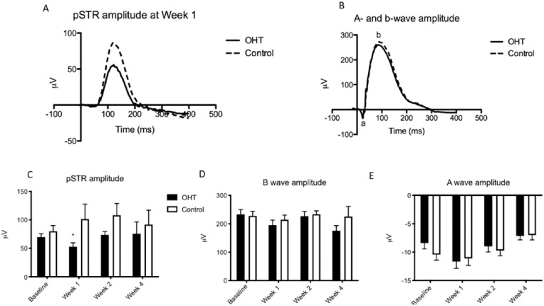
Methods: Adult C57/Bl6 mice (n = 31) were subjected to OHT in one eye using a modified circumlimbal suture technique that elevated intraocular pressure (IOP) for 30 minutes. Contralateral un-operated eyes served as controls. IOP, anterior segment optical coherence tomography, and fundus fluorescein angiography (FFA) were performed. The positive scotopic threshold response (pSTR) and a-wave and b-wave amplitudes were also evaluated. Retinal tissues were immunostained for the retinal ganglion cell (RGC) marker RBPMS and the glial marker GFAP.

Results: OHT eyes developed shallower anterior chambers and dilated pupils. FFA showed focal leakage in 32.2% of OHT eyes, but in none of the control eyes. pSTR was significantly reduced at week 1 in OHT eyes compared to control eyes (57.3 ± 7.2 µV vs. 106.9 ± 24.8 µV; P < 0.05), but a- and b-waves were unaffected. GFAP was upregulated in OHT eyes but not in control eyes or eyes that had been sutured without OHT. RGC density was reduced in OHT eyes after 4 weeks (3857 ± 143.8) vs. control eyes (4469 ± 176.0) (P < 0.05).

Conclusions: Our minimally invasive model resulted in acute OHT with characteristics of AAC in the absence of non-OHT-related neuroinflammatory changes arising from ocular injury alone.

Translational relevance: This model provides a valuable approach to studying specific characteristics of a severe blinding disease in an experimental setting. Focal areas of ischemia were demonstrated, consistent with clinical studies of acute angle closure patients elsewhere, which may indicate the need for further research into how this could affect visual outcome in these patients.

Keywords: acute angle closure; circumlimbal suture; minimally invasive; ocular hypertension.


Conflict of interest statement

Disclosure: R.S. Chong, None; J.M.F. Busoy, None; B. Tan, None; S.W. Yeo, None; Y.S. Lee, None; A.V. Barathi, None; J.G. Crowston, None; L. Schmetterer, None

Figures

Figure 1.
Figure 1.
Experimentally induced acute angle closure resulted in changes in ACD and IOP on the day of surgery. (A, B) AS-OCT images from OHT and control eyes taken intraoperatively show a reduction in ACD in OHT eyes versus controls, marked by double-headed arrows. The iris profile of OHT eyes also appeared steeper, and the pupil diameter was larger in these eyes. (C) H&E staining at week 4 post-induction of acute OHT did not show evidence of peripheral anterior synechiae. (D) ACD measurements were significantly reduced in OHT eyes as compared to control eyes intraoperatively (day 0) with the suture left in situ, but also in measurements taken after the suture was cut at 30 minutes. ACD in OHT versus control eyes did not differ significantly thereafter. (E) Intraoperative IOP measurements showed an initial spike in OHT eyes of up to 80 mm Hg, followed by a gradual reduction until the suture was cut at 30 minutes. (F, G) Cumulative and mean IOP measured intraoperatively with the suture left in situ showed significant increase in the OHT eyes versus controls. **P < 0.01; n = 31.
Figure 2.
Figure 2.
Acute angle closure resulted in a significant reduction in RGC counts and GCC thickness at 4 weeks following induction of raised intraocular pressure. (A, B) Representative images of flatmounted retinas from OHT and control eyes, respectively, with the RGC marker RBPMS in green. Scale bar: 100 µm. (C) GCC thickness was measured from OCT images centered at the optic nerve head, in the area demarcated by double-headed arrows. Scale bar: 100 µm. (D) RGC counts in flatmounted retinas were significantly reduced in OHT eyes versus controls at 4 weeks following induction of acute angle closure. (E) RGC counts in OHT eyes that showed vascular leakage were significantly reduced as compared to eyes that did not show vascular leakage. (F, G) GCC thickness of OHT versus control eyes was also reduced at both 2 and 4 weeks following induction of acute angle closure. (H, I) Total retinal thickness at 2 and 4 weeks did not differ significantly between OHT and control eyes. *P < 0.05, **P < 0.01. Sample size at week 2, n = 26; week 4, n = 2.
Figure 3.
Figure 3.
OHT eyes demonstrated early loss of inner retinal electrophysiological response, but outer retinal responses were preserved. (A, B) Representative pSTR amplitudes and a- and b-wave responses, respectively, in OHT and control eyes. (C) pSTR amplitudes in OHT eyes were significantly reduced at week 1 but subsequently increased again relative to control eyes. (D, E) The b- and a-wave amplitudes, respectively, do not demonstrate any significant difference in OHT versus control eyes at any time point. *P < 0.05. Sample size at baseline, n = 31; week 1, n = 31; week 2, n = 26; week 4, n = 21.
Figure 4.
Figure 4.
Acute angle closure resulted in focal leakage of retinal capillaries in some OHT eyes, which was not seen in controls. (A–C) Baseline fundal photographs, low-resolution FFA, and high-resolution FFA, respectively, for comparison. Fundus photograph at week 1 (D) shows a focal area of hypopigmentation, with associated capillary leakage on low-resolution FFA (E) and high-resolution FFA (F). Gross vessel morphology and retinal perfusion looked otherwise intact. (G, H) Retinal sections from OHT eyes showed variable evidence of GFAP (green) immunostaining at week 1. Contralateral eyes that had been sutured without knotting and did not experience OHT had an appearance similar to that of G.

References

    1. Quigley HA, Broman AT. The number of people with glaucoma worldwide in 2010 and 2020. Br J Ophthalmol. 2006; 90: 253–254. - PMC - PubMed
    1. Friedman DS, Foster PJ, Aung T, He M. Angle closure and angle-closure glaucoma: what we are doing now and what we will be doing in the future. Clin Exp Ophthalmol. 2012; 40: 381–387. - PubMed
    1. Tham YC, Li X, Wong TY, Quigley HA, Aung T, Cheng CY. Global prevalence of glaucoma and projections of glaucoma burden through 2040: a systematic review and meta-analysis. Ophthalmology. 2014; 121: 2081–2090. - PubMed
    1. Aung T, Lim MC, Chan YH, Rojanapongpun P, Chew PT, EXACT Study Group. Configuration of the drainage angle, intraocular pressure, and optic disc cupping in subjects with chronic angle-closure glaucoma. Ophthalmology. 2005; 112: 28–32. - PubMed
    1. Sun X, Dai Y, Chen Y, et al. .. Primary angle closure glaucoma: what we know and what we don't know. Prog Retin Eye Res. 2017; 57: 26–45. - PubMed

Publication types