. 2020 Jun 20;7(6):602-607.
doi: 10.1364/optica.388157. Epub 2020 Jun 4.

Single-molecule orientation localization microscopy for resolving structural heterogeneities between amyloid fibrils

Affiliations

Single-molecule orientation localization microscopy for resolving structural heterogeneities between amyloid fibrils

Tianben Ding et al. Optica. .

Abstract

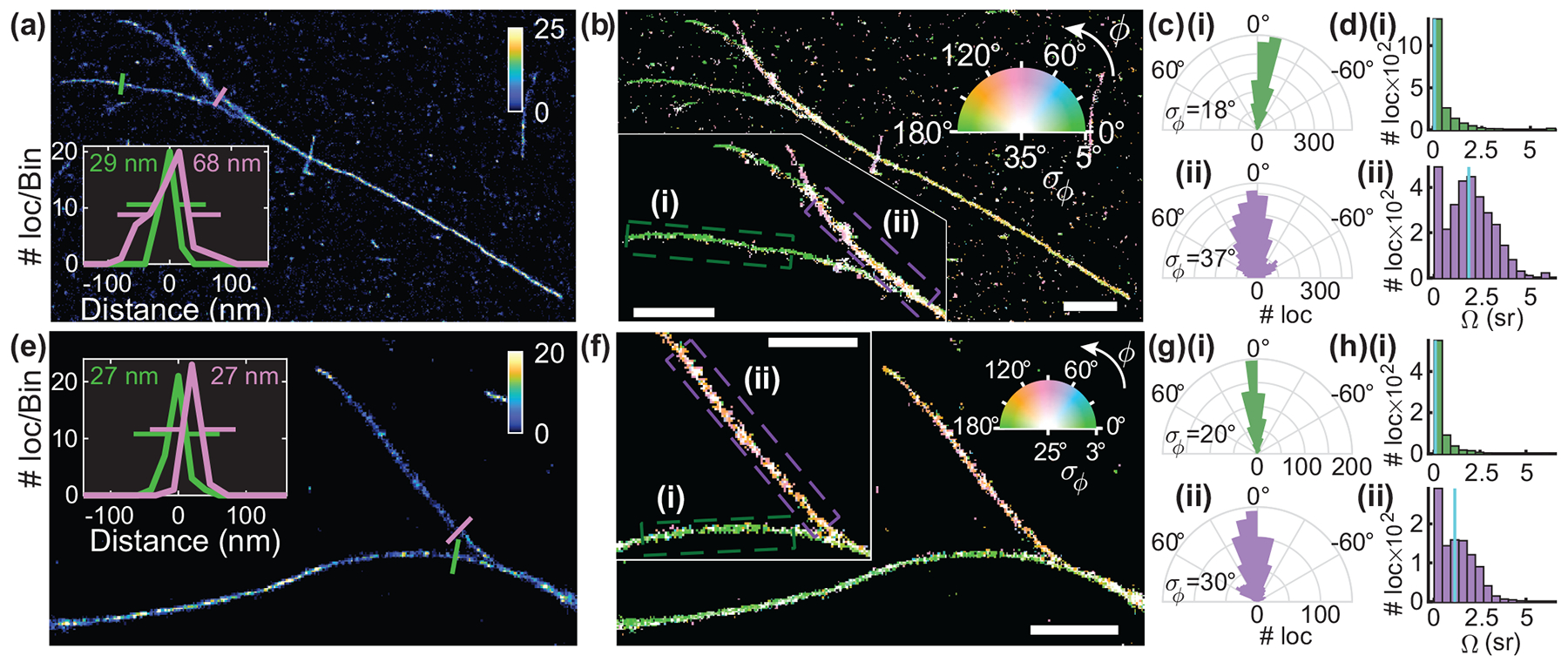
Simultaneous measurements of single-molecule positions and orientations provide critical insight into a variety of biological and chemical processes. Various engineered point spread functions (PSFs) have been introduced for measuring the orientation and rotational diffusion of dipole-like emitters, but the widely used Cramér-Rao bound (CRB) only evaluates performance for one specific orientation at a time. Here, we report a performance metric, termed variance upper bound (VUB), that yields a global maximum CRB for all possible molecular orientations, thereby enabling the measurement performance of any PSF to be computed efficiently (~1000× faster than calculating average CRB). Our VUB reveals that the simple polarized standard PSF provides robust and precise orientation measurements if emitters are near a refractive index interface. Using this PSF, we measure the orientations and positions of Nile red (NR) molecules transiently bound to amyloid aggregates. Our super-resolved images reveal the main binding mode of NR on amyloid fiber surfaces, as well as structural heterogeneities along amyloid fibrillar networks, that cannot be resolved by single-molecule localization alone.


Conflict of interest statement

Disclosures. The authors declare no conflicts of interest.

Figures

Fig. 1.
Fig. 1.
Orientation measurement precision using various point spread functions (PSFs) for 380 signal photons and 2 background photons per pixel. (a) Average (over orientation space) trace RΣ of the CRB R, versus the trace ΓΣ of our variance upper bound (VUB) for various PSFs. Blue: unpolarized standard (std), red: polarized standard (polar), yellow: bifocal microscope (bif), purple: bisected (bi), green: double helix (DH), aqua: tri-spot (tri), and maroon: quadrated (qua) PSF. Dark line: ΓΣ = mean of RΣ. Inset: error of ΓΣ relative to average RΣ. Bars indicate minimum/maximum error relative to the mean (circle) over all of space. (b) Fisher information (FI) lower bound [Γ−1]jj of each orientational second moment mj. (c) Trace ΓΣ of VUB for various refractive indices. Dotted line: refractive index of water. Simulated orthogonally polarized images of (d) a fixed molecule with orientation (θ = 90°, ϕ = 45°) and an isotropic emitter using (e) the polarized PSF and (f) the tri-spot PSF. Insets: normalized noiseless PSF images. Scale bars: 400 nm.
Fig. 2.
Fig. 2.
Transient amyloid binding (TAB) SMLM and SMOLM images acquired using Nile red (NR). (a) SMLM image of a network of Aβ42 fibrils. Color bar: localizations per bin (20 × 20 nm2). Inset: diffraction-limited image. (b) TAB SMOLM image, color-coded according to the mean azimuthal (ϕ) orientation of NR molecules measured within each bin. Inset: Main binding mode of NR to β-sheets, i.e., dipole moments aligned mostly parallel to the long axis of a fibril (its backbone). (c-g) All individual orientation measurements localized along fibril backbones within the white boxes in (b). The lines are oriented and color-coded according to the direction of the estimated ϕ angle. Red dashed lines depict fibril backbones estimated from the SMLM image. Scale bars: (a,b) 1 μm, (f,g) 100 nm. Orientation-localization data are available in Dataset 1 [30].
Fig. 3.
Fig. 3.
Structural heterogeneity of Aβ42 fibrils revealed by TAB SMOLM imaging. (a) SMLM image of fibril bundles. Color bar: localizations per bin (20 × 20 nm2). Inset: fibril cross-sections at the locations denoted by green and purple lines with measured full-width at half-maximum (FWHM) thicknesses. (b) TAB SMOLM image corresponding to (a), color-coded according to the mean (ϕ) and standard deviation (σϕ) of the azimuthal orientation measured within each bin. Inset: zoomed (i) thin and (ii) thick fibril regions isolated from background structures. (c) Histograms of measured azimuthal orientations relative to the fibril backbone within the regions denoted in (b), showing standard deviations (σϕ) of (i) 18° and (ii) 37°. (d) Measured wobbling areas (Ω) corresponding to the localizations in (c), yielding median wobbling areas (Ωmed, cyan) of (i) 0.07 sr and (ii) 1.89 sr. (e-h) TAB SMLM and SMOLM images of another fibril field of view, as in (a-d). Although fibril regions (i) and (ii) in the inset of (f) show little difference in apparent width, the measured orientation distributions contain significant differences. The standard deviations of azimuthal angles σϕ are (i) 20° vs (ii) 30°, and the median wobbling areas (Ωmed, cyan) are (i) 0.07 sr vs (ii) 1.12 sr. Scale bars: 1 μm. Orientation-localization data are available in Dataset 1 [30].

References

    1. Sosa H, Peterman EJ, Moerner WE, and Goldstein LS, “ADP-induced rocking of the kinesin motor domain revealed by single-molecule fluorescence polarization microscopy,” Nat. Struct. Biol 8, 540–544 (2001). - PubMed
    1. Peterman EJ, Sosa H, Goldstein LS, and Moerner W, “Polarized Fluorescence Microscopy of Individual and Many Kinesin Motors Bound to Axonemal Micro-tubules,” Biophys. J 81, 2851–2863 (2001). - PMC - PubMed
    1. Lippert LG, Dadosh T, Hadden JA, Karnawat V, Diroll BT, Murray CB, Holzbaur ELF, Schulten K, Reck-Peterson SL, and Goldman YE, “Angular measurements of the dynein ring reveal a stepping mechanism dependent on a flexible stalk,” Proc. Natl. Acad. Sci 114, E4564–E4573 (2017). - PMC - PubMed
    1. Ha T, Glass J, Enderle T, Chemla DS, and Weiss S, “Hindered Rotational Diffusion and Rotational Jumps of Single Molecules,” Phys. Rev. Lett 80, 2093–2096 (1998).
    1. Backer AS, Biebricher AS, King GA, Wuite GJL, Heller I, and Peterman EJG, “Single-molecule polarization microscopy of DNA intercalators sheds light on the structure of S-DNA,” Sci. Adv 5, eaav1083 (2019). - PMC - PubMed

LinkOut - more resources