

Synthesis of the Novel AT1 Receptor Tracer [18F]Fluoropyridine-Candesartan via Click Chemistry
- PMID: 32832788
- PMCID: PMC7439361
- DOI: 10.1021/acsomega.0c02310
Synthesis of the Novel AT1 Receptor Tracer [18F]Fluoropyridine-Candesartan via Click Chemistry
Abstract
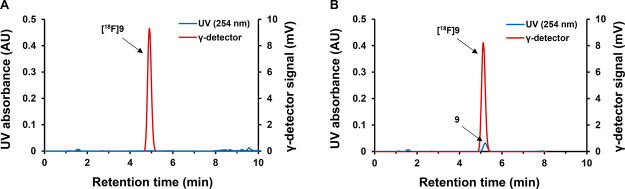
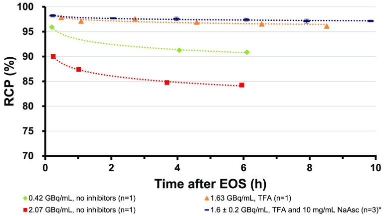
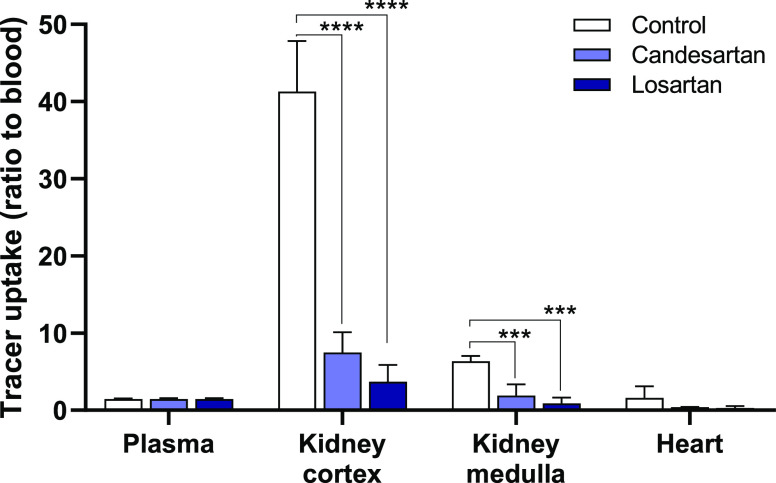
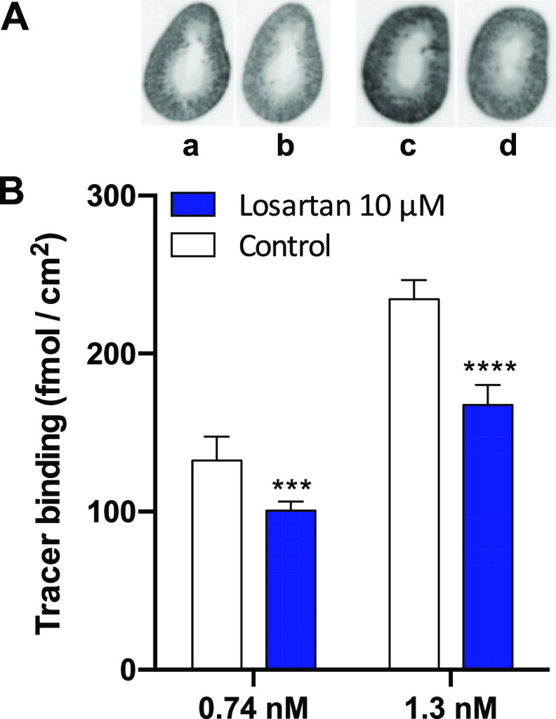
A novel 7-((4-(3-((2-[18F]fluoropyridin-3-yl)oxy)propyl)-1H-1,2,3-triazol-1-yl)methyl)-1H-benzo[d]imidazole derivative of the angiotensin II type-1 receptor (AT1R) blocker candesartan, [18F]fluoropyridine-candesartan, was synthesized via the copper-catalyzed azide-alkyne cycloaddition click reaction between 2-[18F]fluoro-3-(pent-4-yn-1-yloxy)pyridine ([18F]FPyKYNE) and the tetrazole-protected azido-candesartan derivative, followed by acid deprotection. This three-step, two-pot, and two-step purification synthesis was done within 2 h. The use of tris[(1-hydroxypropyl-1H-1,2,3-triazol-4-yl)methyl]amine (THPTA) as a Cu(I) stabilizing agent increased the overall radiochemical yield by 4-fold (10 ± 2%, n = 13) compared to the reaction without THPTA (2.4 ± 0.2%, n = 3; decay-corrected from 18F produced at the end-of-beam). Complete separation of [18F]FPyKYNE from its nitro precursor and [18F]fluoropyridine-candesartan from the deprotected azido-candesartan allowed for high molar activities (>380 GBq/μmol) of the tracer. The use of 0.1% trifluoroacetic acid in water for reformulation and the addition of sodium ascorbate to the final formulation (1.6 ± 0.2 GBq/mL, n = 3) prevented tracer radiolysis with >97% radiochemical purity for a period of up to 10 h after the end-of-synthesis. A significant reduction in the uptake (86 ± 3%, n = 8) of the tracer was observed ex vivo in rats (at 20 min postinjection) in the AT1R-rich kidney cortex following pretreatment with saturating doses of the AT1R antagonist candesartan or losartan. This specific binding to AT1R was confirmed in vitro in the rat renal cortex (autoradiography) by a reduction of 26 ± 5% (n = 12) with losartan coincubation (10 μM). These favorable binding properties support further studies to assess the potential of [18F]fluoropyridine-candesartan as a tracer for the positron emission tomography imaging of renal AT1R.
Copyright © 2020 American Chemical Society.
Conflict of interest statement
The authors declare no competing financial interest.
Figures
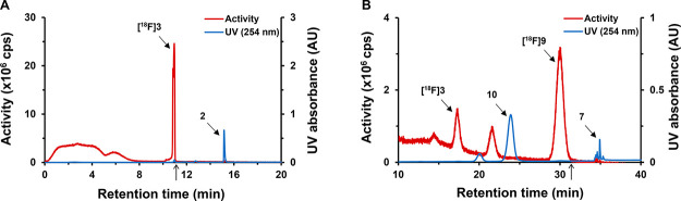
 , following [18F]3 collection). (B)
C18 Column: [18F]fluoropyridine–candesartan ([18F]9) (tR = 30 min)
purification, [18F]3 (tR = 17.5 min), 10 (tR = 24 min), and 7 (tR = 3.5 min, eluted with 100% EtOH after switch
, following [18F]3 collection). (B)
C18 Column: [18F]fluoropyridine–candesartan ([18F]9) (tR = 30 min)
purification, [18F]3 (tR = 17.5 min), 10 (tR = 24 min), and 7 (tR = 3.5 min, eluted with 100% EtOH after switch  , following [18F]9 collection).
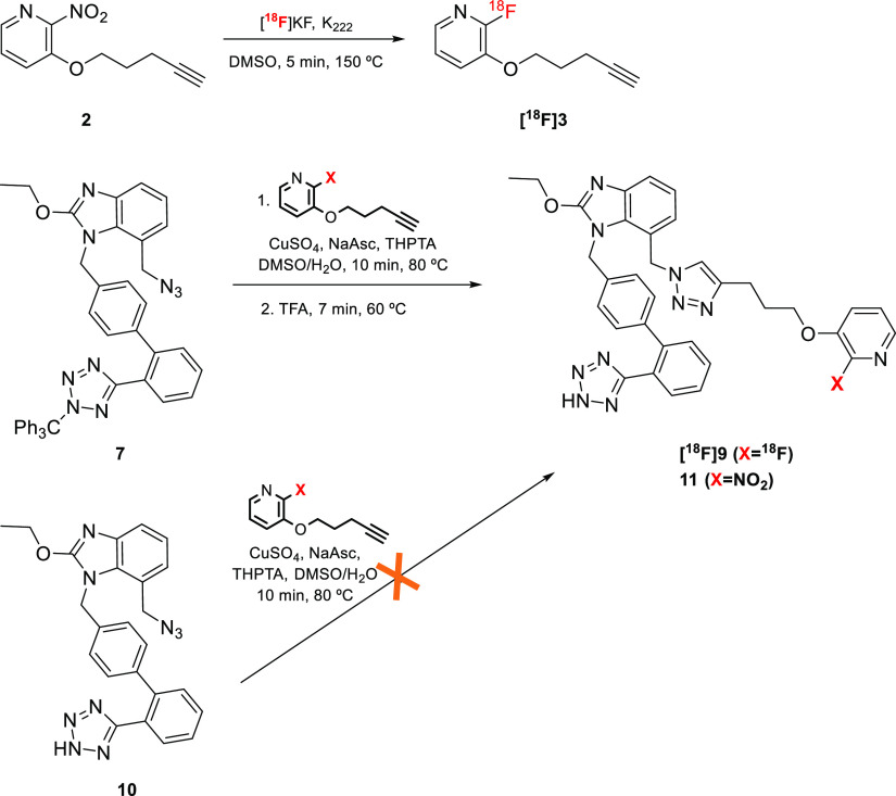
References
-
- de Gasparo M.; Catt K. J.; Inagami T.; Wright J. W.; Unger T. International union of pharmacology. XXIII. The angiotensin II receptors. Pharmacol. Rev. 2000, 52, 415–472. - PubMed
LinkOut - more resources
Full Text Sources
Research Materials
Miscellaneous
