. 2020 Sep 25;48(17):9681-9693.
doi: 10.1093/nar/gkaa694.

A CRISPR-associated factor Csa3a regulates DNA damage repair in Crenarchaeon Sulfolobus islandicus

Affiliations

A CRISPR-associated factor Csa3a regulates DNA damage repair in Crenarchaeon Sulfolobus islandicus

Zhenzhen Liu et al. Nucleic Acids Res. .

Abstract

CRISPR-Cas system provides acquired immunity against invasive genetic elements in prokaryotes. In both bacteria and archaea, transcriptional factors play important roles in regulation of CRISPR adaptation and interference. In the model Crenarchaeon Sulfolobus islandicus, a CRISPR-associated factor Csa3a triggers CRISPR adaptation and activates CRISPR RNA transcription for the immunity. However, regulation of DNA repair systems for repairing the genomic DNA damages caused by the CRISPR self-immunity is less understood. Here, according to the transcriptome and reporter gene data, we found that deletion of the csa3a gene down-regulated the DNA damage response (DDR) genes, including the ups and ced genes. Furthermore, in vitro analyses demonstrated that Csa3a specifically bound the DDR gene promoters. Microscopic analysis showed that deletion of csa3a significantly inhibited DNA damage-induced cell aggregation. Moreover, the flow cytometry study and survival rate analysis revealed that the csa3a deletion strain was more sensitive to the DNA-damaging reagent. Importantly, CRISPR self-targeting and DNA transfer experiments revealed that Csa3a was involved in regulating Ups- and Ced-mediated repair of CRISPR-damaged host genomic DNA. These results explain the interplay between Csa3a functions in activating CRISPR adaptation and DNA repair systems, and expands our understanding of the lost link between CRISPR self-immunity and genome stability.


Figures

Figure 1.
Figure 1.
Organization of CRISPR adaptation module (A), ups (B) and ced (C) operons in S. islandicus REY15A. The adaptation module encodes four genes essential for CRISPR adaptation of subtype I-A system. Diamonds and rectangles in the CRISPR arrays represent repeats and spacers; respectively, 115 and 93 are the total numbers of spacers in each CRISPR array. The ups cluster: upsX, encodes a protein with unknown function; upsE, encodes a secretion ATPase; upsF, encodes an integral membrane protein; and upsA and upsB, encode two pili subunits. The ced cluster: cedA1 and cedA2: encode two small transmembrane proteins; cedA: encodes a larger transmembrane protein; cedB: encodes a HerA/VirB4 homolog.
Figure 2.
Figure 2.
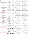
Analysis of Csa3a binding with the promoters of ups, ced, tfb3 and cdc6–2 genes. (A) EMSA analysis of Csa3a binding to the upsX (SiRe_1878), upsE (SiRe_1879), upsA (SiRe_1881), cedA1 (SiRe 1316), cedB (SiRe_1857), tfb3 (SiRe_1717) and cdc6–2 (SiRe_1231) promoters. For binding assays using the wild-type promoters or the mutated promoters, each reaction contained the 5′-end HEX labeled probes: 5 ng/μlL, poly(dI-dC): 5 ng/μl, and Csa3a protein: 40, 80 or 120 ng/μl. For the specific competition assay, each reaction contained the 5′-end HEX labeled probes: 5 ng/μl, poly(dI-dC): 5 ng/μL, and Csa3a protein: 120 ng/μL, and unlabeled specific competitor (cold probe): 10 or 20 ng/μL. The probe location and mutated region on each promoter are indicated in relation to the ATG codon of each open reading frame. The wild-type and mutated sequences are indicated and the UV-responsive element (26) is boxed. P and PM: the wild-type probes and the mutated probes used in EMSA and LSPR experiments, respectively. W: precipitation at loading wells; S: shift; F: free probe. (B) LSPR analysis of fixed Csa3a protein on the chip to bind the promoters and their mutants used in (A). The concentrations of probes used for the analysis are shown. KD = mean±standard derivations of three independent experiments.
Figure 3.
Figure 3.
Analysis of promoter activities using the reporter gene system in the strains with or without csa3a gene. (A) The reporter plasmid used in this study. Promoters of DDR genes were cloned immediately upstream of lacS gene which encodes β-galactosidase. (B) The specific β-galactosidase activities for the tested promoters in wild-type (S. islandicus E233S) or csa3a deletion strains with or without treatment with 2 μM NQO for 6 h. Error bars: standard derivations of three independent experiments. Statistical significance: *P< 0.05, ***P< 0.001, two-way ANOVA and Dunnett.
Figure 4.
Figure 4.
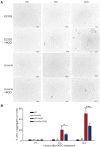
Deletion of csa3a gene reduced DNA damage-induced cell aggregation. (A) Microscopic analysis of cell aggregates in samples taken from the cultures of the wild-type (E233S) and Δcsa3a mutants at different time points with or without NQO treatment. Red arrows indicate the example of cell aggregates, and the aggregate with more than 30 cells in the E233S sample at 12 h post NQO treatment was circled. (B) Quantification data of cell aggregates in the cell samples shown in panel A. At least 1000 cells were analyzed for each sample. Error bars: standard derivations of three independent experiments. Statistical significance: *P< 0.05, ***P< 0.001, two-way ANOVA and Dunnett.
Figure 5.
Figure 5.
The csa3a deleted strain showed higher sensitivity to NQO. (A) Cell cycle profiles of wild-type, Δcsa3a and Δcdc6-2 cultures. DNA contents were divided into 256 arbitrary points on the X-axis, and cell counts (Y-axis) were obtained for each point and plotted against the DNA content. Each sample was grown in SCVU in the presence (denoted as +NQO), or absence (–NQO) of 2 μM NQO for 6 h. DNA-less cells (L); cells containing one chromosome (1), and cells containing two chromosomes (2). Red arrows indicate the DNA-less cells. (B) Growth curves based on absorbance at 600 nm. (C) Plate titration of cells. Each strain was grown in the absence or presence of NQO for 48 h, and a series of dilutions were prepared for each sample which was plated for 12 h. WT: S. islandicus E233S; Δcas3a: csa3a deletion strain; Δcdc6-2: cdc6-2 deletion strain. Error bars: standard derivations of three independent experiments. (D) Survival rates of S. islandicus wild-type (E233S) and Δcsa3a strains after NQO treatment. Exponentially growing strains were treated with 2 μM NQO for 6 h. Cell samples were plated on NQO-free SCVYU plates for determination of colony formation units (CFU)/mL culture. Survival rate = (CFU/mL of NQO treated cells)/(CFU/ml of non-treated cells). Statistical significance: ***P< 0.001, two-way ANOVA and Dunnett.
Figure 6.
Figure 6.
Deletion of csa3a gene reduced the efficiency of Ups- and Ced-mediated repair of CRISPR-damaged host genomic DNA. (A) schematic diagram of Ups- and Ced-mediated DNA repair analysis. Plasmid pTcas5 carrying a mini-CRISPR (Repeat-Spacer-Repeat) with the spacer against cas5 gene was electroporated into S. islandicus wt (ΔpyrEFΔlacS) and Δcsa3apyrEFΔlacSΔcsa3a) cells (step 1). After electroporation, the transformed cells were incubated with or without Δcas5pyrEFΔlacSΔcas5) mating partner cells for 2 h at 78°C (step 2) and then plated in the plates of SCV medium without uracil (step 3). (B) schematic diagram of homologous recombination of CRISPR-damaged genomic DNA in wt or Δcsa3a cells carrying the pTcas5 self-targeting plasmid and containing the donor DNA imported from Δcas5 cells. Subtype I-A Cascade complex targeted site at cas5 gene locus is shown in red. The cas5 gene indicates as a blue arrow. Deletion of the cas5 gene is indicated as a dash line. Double-crossover between the homologous regions upstream and downstream of cas5 gene is shown. F and R represent the primers used for PCR amplification of the cas5 gene locus on the chromosome of the colonies. (C) fold increases of the transformation efficiencies of wt or Δcsa3a cells incubated with Δcas5 mating partner cells compared with that of wt or Δcsa3a cells (E[wt::pTcas5×Δcas5]/E[wt::pTcas5] or Ecsa3a::pTcas5×Δcas5]/Ecsa3a::pTcas5]; E: transformation efficiency) were calculated, respectively. Statistical significance: *P< 0.05, two-way ANOVA and Dunnett. (D) PCR amplification of the cas5 gene locus on the chromosomes of 20 randomly selected wt::pTcas5 × Δcas5, Δcsa3a::pTcas5 × Δcas5, wt::pTcas5 and Δcsa3a::pTcas5 single colonies, respectively, on SCV plates without addition of uracil. L, DNA ladder; a, PCR control using S. islandicus REY15A genomic DNA as the template; b, PCR control using the Δcas5 strain genomic DNA as the template; 1–20, PCR amplification of cas5 gene locus using 20 randomly selected colonies, respectively.
Figure 7.
Figure 7.
A proposal for Csa3a-centered network of DNA damage response for CRISPR immunity in S. islandicus. MGE invasion activates the expression of the global regulator Csa3a (32). Csa3a in turn activates expression of DDR genes, including ups and ced operons, and triggers CRISPR adaptation and immunity against invaders (7,8). However, a subset of host DNA derived spacers would guide self-immunity against the genomic DNA. The activated Ups system aggregates cells and the Ced system transfers DNA from adhered cells into the target cells. The transferred DNA was used as the donor DNA for homologous recombination (HR) to remove newly integrated spacers and to repair the DNA breaks, resulting in intact genomic DNA. Ups: UV-responsive pili of Sulfolobus; Ced: Crenarchaeal system for exchange of DNA. Diamond: the CRISPR repeat sequence.

Similar articles

Cited by

References

    1. Barrangou R., Fremaux C., Deveau H., Richards M., Boyaval P., Moineau S., Romero D.A., Horvath P.. CRISPR provides acquired resistance against viruses in prokaryotes. Science. 2007; 315:1709–1712. - PubMed
    1. Makarova K.S., Haft D.H., Barrangou R., Brouns S.J., Charpentier E., Horvath P., Moineau S., Mojica F.J., Wolf Y.I., Yakunin A.F. et al. .. Evolution and classification of the CRISPR−Cas systems. Nat. Rev. Microbiol. 2011; 9:467–477. - PMC - PubMed
    1. Koonin E.V., Makarova K.S., Zhang F.. Diversity, classification and evolution of CRISPR−Cas systems. Curr. Opin. Microbiol. 2017; 37:67–78. - PMC - PubMed
    1. Charpentier E., Richter H., van der Oost J., White M.F.. Biogenesis pathways of RNA guides in archaeal and bacterial CRISPR−Cas adaptive immunity. FEMS Microbiol. Rev. 2015; 39:428–441. - PMC - PubMed
    1. Plagens A., Richter H., Charpentier E., Randau L.. DNA and RNA interference mechanisms by CRISPR−Cas surveillance complexes. FEMS Microbiol. Rev. 2015; 39:442–463. - PMC - PubMed

Publication types