

doi: 10.1164/rccm.202007-2676LE.
Injury to the Endothelial Glycocalyx in Critically Ill Patients with COVID-19
Affiliations
- PMID: 32833500
- PMCID: PMC7560808
- DOI: 10.1164/rccm.202007-2676LE
Item in Clipboard
Injury to the Endothelial Glycocalyx in Critically Ill Patients with COVID-19
Am J Respir Crit Care Med.
.
No abstract available
Figures
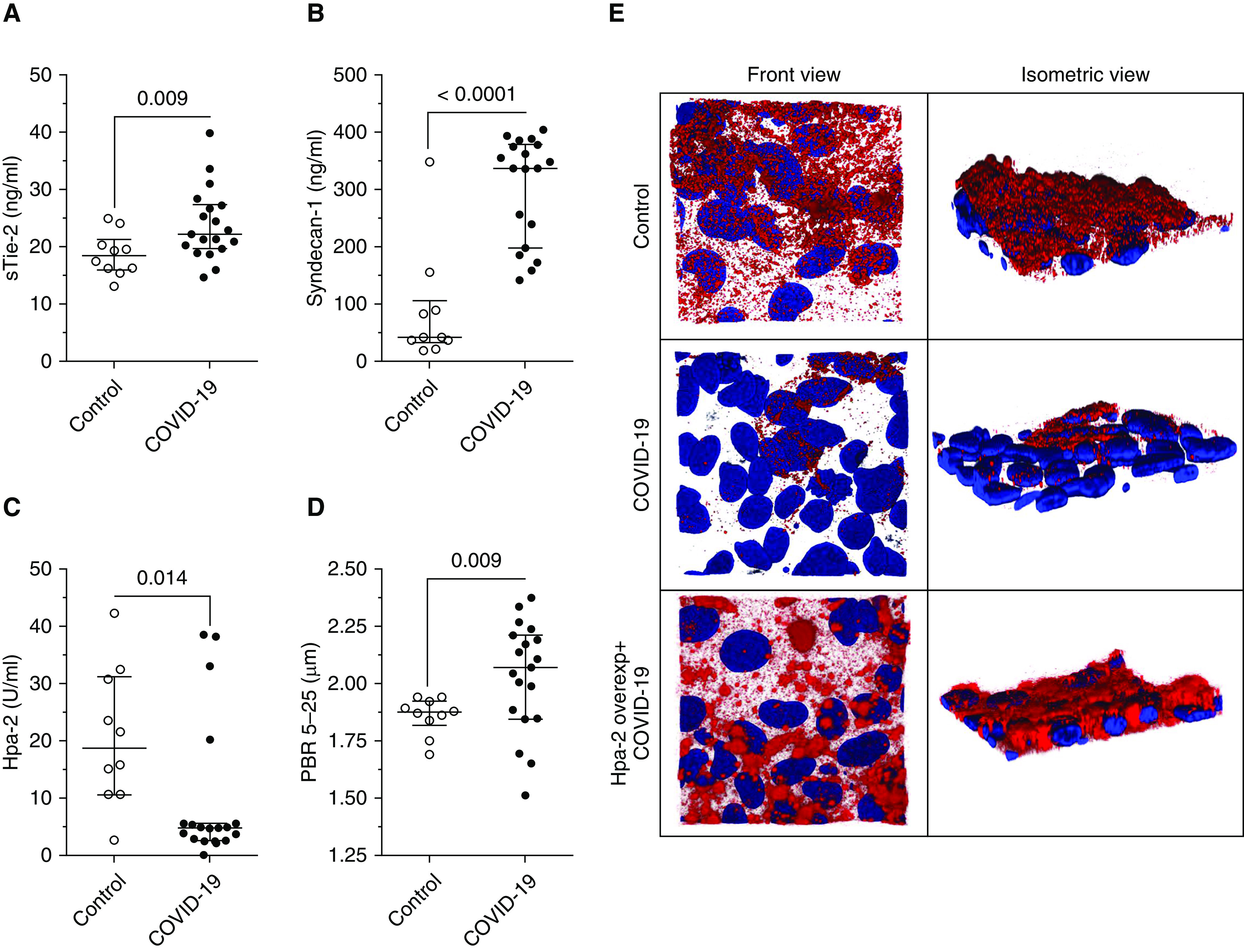
Injury to the endothelial glycocalyx in severe coronavirus disease (COVID-19). Scatter dot plots (median [interquartile range]) show (A) sTie-2 and (B) syndecan-1 concentrations for control and patients with COVID-19. Concentrations for both syndecan-1 (control: 41.5 [32.6–105.4] ng/ml vs. COVID-19: 336.5 [196.7–377.1] ng/ml) and sTie-2 (control: 18.4 [16–21.3] ng/ml vs COVID-19: 22.3 [19.7–27.3] ng/ml) were increased in patients with COVID-19. (C) Although both Hpa-1 (heparanase-1) concentration and activity were not significantly altered in comparison with control subjects, patients with COVID-19 showed an acquired deficiency in Hpa-2 (heparanase-2) (control: 18.7 [10.6–31.1] U/ml vs. COVID-19: 4.7 [2.6–5.1] U/ml). Consequentially, the Hpa-1:Hpa-2 ratio was higher in patients with COVID-19 (control: 0.08 [0.05–0.17] ng/U vs. COVID-19: 0.35 [0.27–0.66] ng/U). (D) Sidestream darkfield imaging in patients allows the quantification of endothelial glycocalyx thickness as indicated by increased perfused boundary region; this was increased in patients with COVID-19 (control: 1.9 [1.8–1.9] μm vs. COVID-19: 2.1 [1.8–2.2] μm), indicating reduced endothelial glycocalyx thickness. For comparison of groups, first, the normal distribution (D’Agostino-Pearson omnibus and Shapiro-Wilk normality test) of variables was tested, and then (A and D) two-sided unpaired t tests and (B and C) Mann-Whitney tests were used accordingly. (E) Exemplary three-dimensional reconstruction of the heparan sulfate (HS) layer images of naive endothelial cells in a microfluidic chip (HS in red; DAPI nuclei staining in blue) after perfusion with serum of a patient with COVID-19 (middle row) compared with a healthy control subject (upper row) in both front and isometric view angles demonstrates the diffuse loss of HS-rich glycocalyx layer in cells treated with COVID-19 serum. In Hpa-2–overexpressing endothelial cells (lower row), the HS surface layer is protected from injurious effects by perfusion with COVID-19 serum. overexp = overexpressing; PBR = perfused boundary region; sTie-2 = soluble Tie-2.
References
Publication types
MeSH terms
Substances
LinkOut - more resources
Full Text Sources
Other Literature Sources
