. 2020 Aug 24;15(8):e0235898.
doi: 10.1371/journal.pone.0235898. eCollection 2020.

Myo/Nog cells are nonprofessional phagocytes

Affiliations

Myo/Nog cells are nonprofessional phagocytes

Jacquelyn Gerhart et al. PLoS One. .

Abstract

Myo/Nog cells were discovered in the chick embryo epiblast. Their expression of MyoD reflects a commitment to the skeletal muscle lineage and capacity to differentiate into myofibroblasts. Release of Noggin by Myo/Nog cells is essential for normal morphogenesis. Myo/Nog cells rapidly respond to wounding in the skin and eyes. In this report, we present evidence suggesting that Myo/Nog cells phagocytose tattoo ink in tissue sections of human skin and engulf cell corpses in cultures of anterior human lens tissue and magnetic beads injected into the anterior chamber of mice in vivo. Myo/Nog cells are distinct from macrophages in the skin and eyes indicated by the absence of labeling with an antibody to ionized calcium binding adaptor molecule 1. In addition to their primary roles as regulators of BMP signaling and progenitors of myofibroblasts, Myo/Nog cells behave as nonprofessional phagocytes defined as cells whose primary functions are unrelated to phagocytosis but are capable of engulfment.


Conflict of interest statement

There are no competing interests related to this manuscript.

Figures
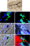
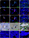
Fig 1
Fig 1. Myo/Nog cells contain ink in human tattooed skin.
An unstained tissue section of tattooed skin is shown in A. Other sections were labeled with the G8 mAb (green) and Hoechst dye (blue). Confocal 60x, 0.5 μM step size images of the same field are shown in B-D. E-F were photographed with the 60x epifluorescent microscope. Green and blue photomicrographs were merged in B and E. Fluorescent photomicrographs were merged with the corresponding DIC image to visualize ink (C, D and F). Tattoo ink appears black in DIC images (C, D and F). Red ink fluoresces in the rhodamine channel of the epifluorescence microscope (G). Some G8+ Myo/Nog cells appeared to contain ink. Bar = 540 μM in A and 5.6 μM in B-G.
Fig 2
Fig 2. Myo/Nog cells contain ink in human tattooed skin.
Tissue sections of tattooed skin were double labeled with the G8 mAb and antibodies to Noggin (Nog) (A-D), MyoD (F-I) or α-SMA (K-N). The colors of the secondary antibodies are indicated in the photographs. Nuclei were stained with Hoechst dye (blue). Images were produced with the epifluorescence (A-E, J and O) and confocal microscopes (F-I and K-N) with 60x lenses. Overlap of green and red fluorescence appears yellow in merged images (C, D, H, I, M and N). Fluorescent photomicrographs were merged with the corresponding DIC image to visualize the ink (black in D, I and N). Some double labeled cells appeared to contain ink (arrows in D, I and N). All ink laden G8+ cells were α-SMA+ (N). Smooth muscle cells of blood vessels also contained α-SMA (K). Minimal to no background fluorescence was visible after staining with the anti-goat (E), anti-IgM and anti-IgG (I), and anti-rabbit (O) secondary antibodies only. Bar = 28 μM in E and 5.6 μM in A-D and G-O.
Fig 3
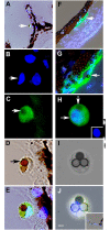
Fig 3. Phagocytosis of dead cells by Myo/Nog cells in human lens tissue.
Anterior lens tissue removed during cataract surgery was treated with doxorubicin then incubated with pHrodo Red that fluoresces after phagocytosis and internalization into acidic compartments of cell. Cells were dissociated and added to untreated lens tissue. A-E and F-I are photographs of two cultures that were fixed and labeled with the G8 mAb (green) and Hoechst dye (blue) 18 hours after addition of treated cells. In K-O and P-S, living cells in untreated lens explants were labeled with the G8 mAb before adding cells from explants treated with doxorubicin and pHrodo Red. Tissue was imaged with the epifluorescence microscope and the 100x (A-I and K-S) or 60x lens (J and T). Unmerged images of nuclei are shown in A, F, K and P. Arrows demonstrate the presence of two nuclei (A and F) or nuclear remnants (P) in a single cell. Merged images of G8 and Hoechst, and pHrodo Red and Hoechst (arrows) are shown in B, G, L and Q, and C, H, M and R, respectively. D, I, N and S are triple merges of blue, green and red. Overlap of red and green appears yellow. Doxorubicin appears black in the quadruple merge of the three fluorochromes and DIC in E, triple merge in I and Hoechst staining and DIC in O (arrows). Some G8+ contained two nuclei or nuclear remnants, pHrodo Red fluorescence (A, I, N and S) and doxorubicin (E and O), indicating phagocytosis. Control cultures labeled with only the secondary antibody and Hoechst dye before (J) or after addition of treated cells (T) illustrate the absence of background green fluorescence. pHrodo Red is visible at the arrow in T. Bar = 9 μm in J and Tand 15 μm in A-I and K-S.
Fig 4
Fig 4. Engulfment of beads by Myo/Nog cells in the anterior cavity.
Beads were injected into the anterior chamber of mice and drawn to the iridocorneal angel with a magnet. One month later, tissue sections were labeled with Hoechst dye (blue) and the G8 mAb (green) and imaged with the epifluorescent microscope and 60x lens. A-E, F and G, and H-J are photographs of bead laden cells on the surface of a ciliary process, within the iridocorneal angle and between the ciliary body and lens, respectively. Cells at the arrows in the merged images of fluorescence and DIC in A and F are shown in B-E and G, respectively. Pigment appears black when photographed with DIC optics (A, D, E and F). Beads (brown/gray) are visible in the DIC images (D and I) and triple merges of DIC, Hoechst dye and G8 (A, E, F, G and J). Arrows point to nuclei in B and H and beads in C, D, G and H. The inset in H shows the Hoechst staining of the nucleus. The inset in J is a section of the ciliary body labeled with the anti-IgM secondary antibody only. Beads are present in the cytoplasm of G8+ Myo/Nog cells. Bar = 9 μm A and F and 2.5 μm in B-E and G-J.
Fig 5
Fig 5. The G8 and Iba1 mAbs label separate populations of cells in the skin and anterior cavity of the eye.
Tissue sections of human tattooed skin (A-D), human lens tissue (E) and sections of the rabbit eye two days after cataract surgery (G-L) were double labeled with the G8 and Iba1 mAbs and imaged with the epifluorescent microscope and 60x lens. The colors of the secondary antibodies are indicated in the photographs. A section through the anterior cavity was stained with hematoxylin and eosin (F). Photographs in B, D and H are quadruple merges of DIC showing black ink and blue, red or green fluorescence. G8+ cells lacked staining for Iba1 in the skin (A and B). Iba1+/G8- cells are shown in a different field in C and D. G8+ cells were present in the human (E) and rabbit lens (G and H), but no Iba1+ cells were detected in these tissues. The arrow in H illustrates the lens capsule. The G8 and Iba1 mAbs labeled separate subpopulations of cells in the ciliary body (cb) (I) and cornea (J). The arrow in J points to an Iba1+ cell. Minimal fluorescence was present in sections of the lens (K) and ciliary body (L) labeled with secondary antibodies only. Bar = 9 μM in A-E and G-L, and 270 μM in F.

References

    1. Gerhart J, Baytion M, DeLuca S, Getts R, Lopez C, Niewenhuis R, et al. DNA dendrimers localize MyoD mRNA in presomitic tissues of the chick embryo. J Cell Biol. 2000;149(4):825–34. Epub 2000/05/17. 10.1083/jcb.149.4.825 . - DOI - PMC - PubMed
    1. Strony R, Gerhart J, Tornambe D, Perlman J, Neely C, Dare J, et al. NeuroM and MyoD are expressed in separate subpopulations of cells in the pregastrulating epiblast. Gene Expr Patterns. 2005;5(3):387–95. Epub 2005/01/22. 10.1016/j.modgep.2004.09.006 . - DOI - PubMed
    1. Gerhart J, Elder J, Neely C, Schure J, Kvist T, Knudsen K, et al. MyoD-positive epiblast cells regulate skeletal muscle differentiation in the embryo. J Cell Biol. 2006;175(2):283–92. Epub 2006/10/25. 10.1083/jcb.200605037 . - DOI - PMC - PubMed
    1. Gerhart J, Neely C, Elder J, Pfautz J, Perlman J, Narciso L, et al. Cells that express MyoD mRNA in the epiblast are stably committed to the skeletal muscle lineage. J Cell Biol. 2007;178(4):649–60. Epub 2007/08/19. 10.1083/jcb.200703060 . - DOI - PMC - PubMed
    1. Gerhart J, Bast B, Neely C, Iem S, Amegbe P, Niewenhuis R, et al. MyoD-positive myoblasts are present in mature fetal organs lacking skeletal muscle. J Cell Biol. 2001;155(3):381–92. Epub 2001/10/31. 10.1083/jcb.200105139 . - DOI - PMC - PubMed

Publication types

MeSH terms