

Post-MI Ventricular Septal Defect During the COVID-19 Pandemic
- PMID: 32835262
- PMCID: PMC7311914
- DOI: 10.1016/j.jaccas.2020.06.019
Post-MI Ventricular Septal Defect During the COVID-19 Pandemic
Abstract
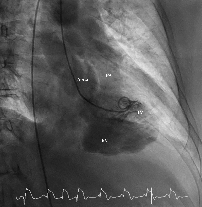
With the COVID-19 pandemic, the fear among patients of contracting it has made them reluctant to seek medical attention on a timely basis even for emergent conditions. We present a case of post infarction ventricular septal rupture due to delayed presentation as a consequence of the fear of COVID-19. (Level of Difficulty: Intermediate.).
Keywords: CAD, coronary artery disease; LAD, left anterior descending; MI, myocardial infarction; PCI, percutaneous coronary intervention; RCA, right coronary artery; RV, right ventricle; VSR, ventricular septal rupture; chest pain; complication; left-sided catheterization; myocardial infarction; right-sided catheterization; ventricular septal defect.
© 2020 The Authors.
Figures


References
-
- Mubarik A., Iqbal A.M. StatPearls; Treasure Island, FL: 2020. Ventricular Septal Rupture. - PubMed
-
- Singh V., Rodriguez A.P., Bhatt P. Ventricular septal defect complicating ST-elevation myocardial infarctions: a call for action. Am J Med. 2017;130:863.e12. - PubMed
-
- Moreyra A.E., Huang M.S., Wilson A.C. Trends in incidence and mortality rates of ventricular septal rupture during acute myocardial infarction. Am J Cardiol. 2010;106:1095–1100. - PubMed
-
- Jeppsson A., Liden H., Johnsson P., Hartford M., Rådegran K. Surgical repair of post infarction ventricular septal defects: a national experience. Eur J Cardiothorac Surg. 2005;27:216–221. - PubMed
-
- Poulsen S.H., Praestholm M., Munk K., Wierup P., Egeblad H., Nielsen-Kudsk J.E. Ventricular septal rupture complicating acute myocardial infarction: clinical characteristics and contemporary outcome. Ann Thorac Surg. 2008;85:1591–1596. - PubMed
Publication types
LinkOut - more resources
Full Text Sources
Miscellaneous
