

Case Reports
doi: 10.1016/j.jaccas.2020.05.060.
Epub 2020 Jun 5.
SARS-CoV-2 Fulminant Myocarditis
Affiliations
- PMID: 32835276
- PMCID: PMC7274592
- DOI: 10.1016/j.jaccas.2020.05.060
Item in Clipboard
Case Reports
SARS-CoV-2 Fulminant Myocarditis
JACC Case Rep.
.
Abstract
An 18-year-old male without prior medical history developed fulminant myocarditis concomitant to severe COVID-19 pneumonia, which was confirmed using serial cardiac magnetic resonance. This may have important diagnostic, monitoring, and pathogenic implications. (Level of Difficulty: Intermediate.).
Keywords: COVID-19, coronavirus disease-2019; EF, ejection fraction; EGE, early gadolinium enhancement; LGE, late gadolinium enhancement; LV, left ventricle; LVEF, left ventricular ejection fraction; SARS-CoV-19, severe acute respiratory syndrome-coronavirus-2019; cardiac magnetic resonance; coronavirus disease 2019; myocarditis.
© 2020 The Authors.
Figures
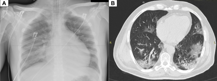
Chest Radiograph and Axial Unenhanced Chest Computed Tomography Scan Obtained on Day 2 After the Onset of Symptoms (A) Chest X-ray and (B) axial unenhanced chest computed tomography scan. Presence of diffuse typical COVID-19–related lesions (typical peripheral opacity, “crazy paving”). COVID-19 = coronavirus disease 2019.
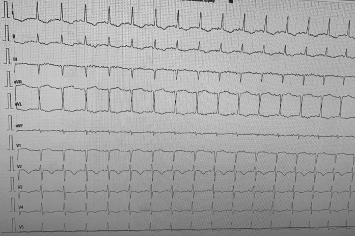
Electrocardiogram on Day 1 Sinus tachycardia (100 beats/min) with negative T waves from V2 to V4.
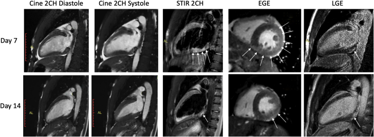
Cardiac Magnetic Resonance: Evolution Between Day 7 and Day 14 Improvement of LV function is shown on cines in the 2CH view, along with a decrease of myocardial edema on T2-weighted STIR (arrows), a decrease of EGE (arrows), and only minor and stable tissue damage on LGE (arrows). 2CH = 2-chamber; EGE = early gadolinium enhancement; LGE = late gadolinium enhancement; LV = left ventricular; STIR = short-tau inversion recovery.
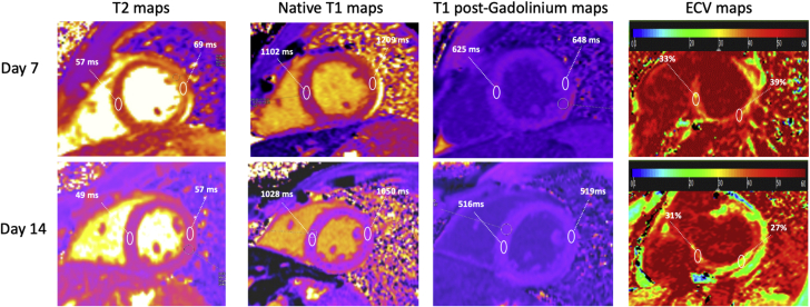
Cardiac Magnetic Resonance: Evolution of T1, T2, and ECVs Between Day 7 and Day 14 Myocardial T2 was increased in the posterolateral wall on Day 7, indicating the presence of edema and it decreased on Day 14. T1 times were higher on Day 7 because of edema. ECV maps depict the dramatic decrease of ECV in the posterolateral wall on Day 14 from 39% to 27%. ECV = extracellular volume.
References
-
- World Health Organization Pneumonia of unknown cause—China. https://www.who.int/csr/don/05-january-2020pneumonia-of-unkown-cause-chi... Available at:
-
- World Health Organization Novel coronavirus—China. https://www.who.int/csr/don/12-january-2020novel-coronavirus-china/en/ Available at:
Publication types
LinkOut - more resources
Full Text Sources
Other Literature Sources
Miscellaneous
