Case Reports
. 2020 Jul 15;2(9):1391-1396.
doi: 10.1016/j.jaccas.2020.05.034. Epub 2020 May 29.

Right Ventricular Clot in Transit in COVID-19: Implications for the Pulmonary Embolism Response Team

Affiliations
Case Reports

Right Ventricular Clot in Transit in COVID-19: Implications for the Pulmonary Embolism Response Team

Sanjum S Sethi et al. JACC Case Rep. .

Abstract

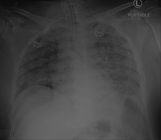
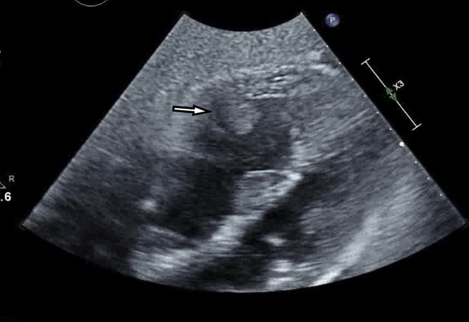
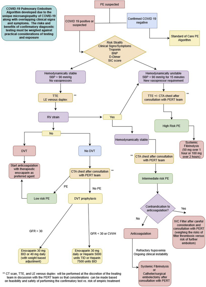
Severe acute respiratory syndrome coronavirus 2 is associated with a prothrombotic state in infected patients. After presenting a case of right ventricular thrombus in a patient with coronavirus disease-2019 (COVID-19), we discuss the unique challenges in the evaluation and treatment of COVID-19 patients, highlighting our COVID-19-modified pulmonary embolism response team algorithm. (Level of Difficulty: Beginner.).

Keywords: ARDS, acute respiratory distress syndrome; COVID-19, coronavirus disease-2019; CTA, computed tomography angiography; ECMO, extracorporeal membrane oxygenation; ICU, intensive care unit; PE, pulmonary embolism; PERT, pulmonary embolism response team; PPE, personal protective equipment; Pao2, partial arterial pressure of oxygen; SARS-CoV-2, severe acute respiratory syndrome-coronavirus-2; TTE, transthoracic echocardiography; VTE, venous thromboembolism; clot in transit; pulmonary embolism; right ventricle; tPA, tissue plasminogen activator; thrombus; vascular disease.


Figures

None
Graphical abstract
Figure 1
Figure 1
Chest Radiograph Demonstrating Bilateral Opacities Indicative of ARDS Anterior posterior portable chest radiograph with fluffy bilateral infiltrates significant for acute respiratory distress syndrome (ARDS) due to coronavirus disease-2019 illness.
Figure 2
Figure 2
Thrombus Visualized in the Mid–Right Ventricle Subcostal view on transthoracic echocardiogram. The right ventricle is enlarged, and there is a thrombus adherent to the free wall of the mid–right ventricle.
Figure 3
Figure 3
Suggested Algorithm for the Diagnosis and Treatment of Venous Thromboembolism in COVID-19 Patients Consensus algorithm determined by members of our institution’s (Columbia University Irving Medical Center) pulmonary embolism response (PERT) team. (Please note: This algorithm is not a societal guideline.) BID = twice daily; BNP = B-type natriuretic peptide; COVID-19 = coronavirus disease-2019; CTA = computed tomography angiography; DVT = deep vein thrombosis; IVC = inferior vena cava; LE = lower extremity; PE = pulmonary embolism; PERT = pulmonary embolism response team; RV = right ventricular; SBP= systolic blood pressure; SIC = sepsis-induced coagulopathy; TID = 3 times daily; TTE = transthoracic echocardiography.

References

    1. Siddiqi H.K., Mehra M. COVID-19 illness in native and immunosuppressed states: a clinical-therapeutic staging proposal. J Heart Lung Transplant. 2020;39:405–407. - PMC - PubMed
    1. Thachil J., Tang N., Gando S. ISTH interim guidance on recognition and management of coagulopathy in COVID-19. J Thromb Haemost. 2020;18:1023–1026. - PMC - PubMed
    1. Bikdeli B., Madhavan M.V., Jimenez D. COVID-19 and thrombotic or thromboembolic disease: implications for prevention, antithrombotic therapy, and follow-up. J Am Coll Cardiol. 2020;75:2950–2973. - PMC - PubMed
    1. Luo W., Yu H., Gou J. Clinical pathology of critical patient with novel coronavirus pneumonia (COVID-19) Preprints. 2020
    1. Ng K.H., Wu A.K., Cheng V.C. Pulmonary artery thrombosis in a patient with severe acute respiratory syndrome. Postgrad Med J. 2005;81:e3. - PMC - PubMed

Publication types