Observational Study
. 2020 May;1(1):e14-e23.
doi: 10.1016/S2666-5247(20)30004-5. Epub 2020 Apr 21.

Comparative tropism, replication kinetics, and cell damage profiling of SARS-CoV-2 and SARS-CoV with implications for clinical manifestations, transmissibility, and laboratory studies of COVID-19: an observational study

Affiliations
Observational Study

Comparative tropism, replication kinetics, and cell damage profiling of SARS-CoV-2 and SARS-CoV with implications for clinical manifestations, transmissibility, and laboratory studies of COVID-19: an observational study

Hin Chu et al. Lancet Microbe. 2020 May.

Abstract

Background: Severe acute respiratory syndrome coronavirus 2 (SARS-CoV-2) was reported from China in January, 2020. SARS-CoV-2 is efficiently transmitted from person to person and, in 2 months, has caused more than 82 000 laboratory-confirmed cases of coronavirus disease 2019 (COVID-19) and 2800 deaths in 46 countries. The total number of cases and deaths has surpassed that of the 2003 severe acute respiratory syndrome coronavirus (SARS-CoV). Although both COVID-19 and severe acute respiratory syndrome (SARS) manifest as pneumonia, COVID-19 is associated with apparently more efficient transmission, fewer cases of diarrhoea, increased mental confusion, and a lower crude fatality rate. However, the underlying virus-host interactive characteristics conferring these observations on transmissibility and clinical manifestations of COVID-19 remain unknown.

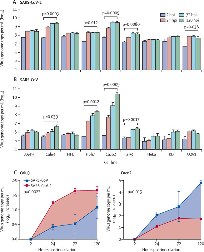
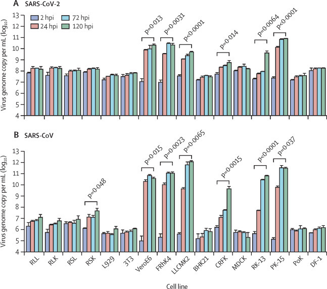
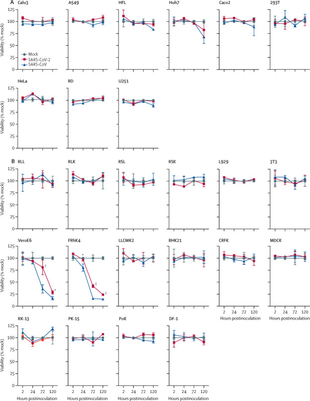
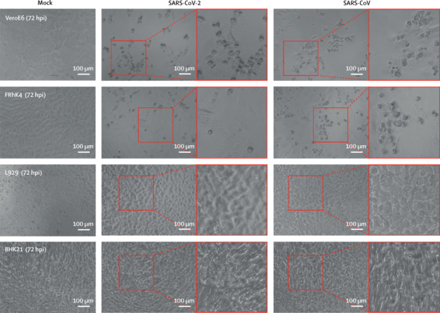
Methods: We systematically investigated the cellular susceptibility, species tropism, replication kinetics, and cell damage of SARS-CoV-2 and compared findings with those for SARS-CoV. We compared SARS-CoV-2 and SARS-CoV replication in different cell lines with one-way ANOVA. For the area under the curve comparison between SARS-CoV-2 and SARS-CoV replication in Calu3 (pulmonary) and Caco2 (intestinal) cells, we used Student's t test. We analysed cell damage induced by SARS-CoV-2 and SARS-CoV with one-way ANOVA.

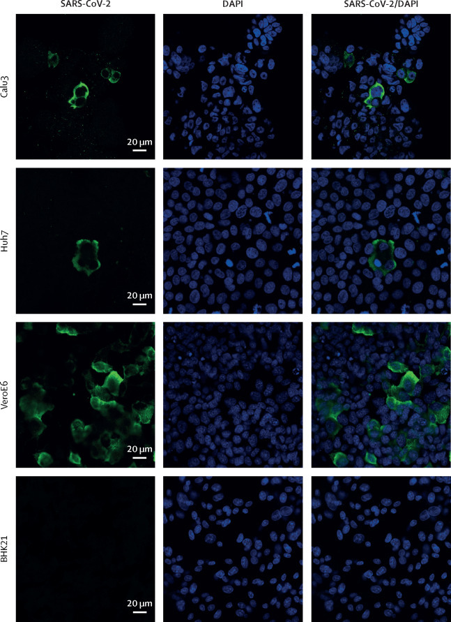
Findings: SARS-CoV-2 infected and replicated to comparable levels in human Caco2 cells and Calu3 cells over a period of 120 h (p=0·52). By contrast, SARS-CoV infected and replicated more efficiently in Caco2 cells than in Calu3 cells under the same multiplicity of infection (p=0·0098). SARS-CoV-2, but not SARS-CoV, replicated modestly in U251 (neuronal) cells (p=0·036). For animal species cell tropism, both SARS-CoV and SARS-CoV-2 replicated in non-human primate, cat, rabbit, and pig cells. SARS-CoV, but not SARS-CoV-2, infected and replicated in Rhinolophus sinicus bat kidney cells. SARS-CoV-2 consistently induced significantly delayed and milder levels of cell damage than did SARS-CoV in non-human primate cells (VeroE6, p=0·016; FRhK4, p=0·0004).

Interpretation: As far as we know, our study presents the first quantitative data for tropism, replication kinetics, and cell damage of SARS-CoV-2. These data provide novel insights into the lower incidence of diarrhoea, decreased disease severity, and reduced mortality in patients with COVID-19, with respect to the pathogenesis and high transmissibility of SARS-CoV-2 compared with SARS-CoV.

Funding: May Tam Mak Mei Yin, The Shaw Foundation Hong Kong, Richard Yu and Carol Yu, Michael Seak-Kan Tong, Respiratory Viral Research Foundation, Hui Ming, Hui Hoy and Chow Sin Lan Charity Fund, Chan Yin Chuen Memorial Charitable Foundation, Marina Man-Wai Lee, The Hong Kong Hainan Commercial Association South China Microbiology Research Fund, The Jessie & George Ho Charitable Foundation, Perfect Shape Medical, The Consultancy Service for Enhancing Laboratory Surveillance of Emerging Infectious Diseases and Research Capability on Antimicrobial Resistance for the Department of Health of the Hong Kong Special Administrative Region Government, The Theme-Based Research Scheme of the Research Grants Council, Sanming Project of Medicine in Shenzhen, and The High Level-Hospital Program, Health Commission of Guangdong Province, China.


Figures

Figure 1
Figure 1
Cell tropism profile of SARS-CoV-2 in human cells originating from different organ tissues Nine cell lines from different human tissues or organs were challenged with SARS-CoV-2 (A) or SARS-CoV (B) at 0·1 MOI. Viral supernatant samples were harvested at 2 hpi, 24 hpi, 72 hpi, and 120 hpi. Viral loads were ascertained with quantitative RT-PCR. For each cell type, the mean viral load at 120 hpi was compared with the mean baseline viral load at 2 hpi. (C) Area under the curve analysis of Calu3 (pulmonary) and Caco2 (intestinal) cells infected with SARS-CoV-2 and SARS-CoV. Bars (A, B) or datapoints (C) represent the mean (error bars show SD) of three independent experiments. Statistical significance was calculated with one-way ANOVA (A, B) or Student's t test (C). SARS-CoV-2=severe acute respiratory syndrome coronavirus 2. SARS-CoV=severe acute respiratory syndrome coronavirus. MOI=multiplicity of infection. hpi=hours postinoculation.
Figure 2
Figure 2
Cell tropism profile of SARS-CoV-2 in non-human cells originating from different animal species 16 non-human cell lines were challenged with SARS-CoV-2 (A) or SARS-CoV (B) at 0·1 MOI. Viral supernatant samples were harvested at 2 hpi, 24 hpi, 72 hpi, and 120 hpi. Viral loads were ascertained with quantitative RT-PCR. For each cell type, the mean viral load at 120 hpi was compared with the mean baseline viral load at 2 hpi. Bars represent the mean (error bars show SD) of three independent experiments. Statistical significance was calculated with one-way ANOVA. SARS-CoV-2=severe acute respiratory syndrome coronavirus 2. SARS-CoV=severe acute respiratory syndrome coronavirus. MOI=multiplicity of infection. hpi=hours postinoculation.
Figure 3
Figure 3
Cell viability profile of SARS-CoV-2-inoculated and SARS-CoV-inoculated cells The cell viability of nine human cell lines (A) and 16 non-human cell lines (B) on SARS-CoV-2 or SARS-CoV infection at 0·1 MOI was quantified at 2 hpi, 24 hpi, 72 hpi, and 120 hpi. For VeroE6 and FRhK4 cells, the mean cell viability of SARS-CoV-2-inoculated cells at each timepoint was compared with that of SARS-CoV-inoculated cells. Datapoints represent the mean (error bars show SD) of three independent experiments. Statistical significance between groups was calculated with one-way ANOVA. SARS-CoV-2=severe acute respiratory syndrome coronavirus 2. SARS-CoV=severe acute respiratory syndrome coronavirus. MOI=multiplicity of infection. hpi=hours postinoculation. *p=0·013. †p=0·0044. ‡p=0·0008.
Figure 4
Figure 4
Detection of SARS-CoV-2-induced cytopathic effects in representative cell types SARS-CoV-2-induced cytopathic effects were assessed in VeroE6 and FRhK4 cells. L929 and BHK21 cells were included as negative controls. Cells were infected with SARS-CoV-2 or SARS-CoV at 0·1 MOI. At 72 hpi, typical cytopathic effects were seen, including cell rounding, detachment, degeneration, and syncytium formation. Boxed area is shown adjacent to each image. Cells were imaged with a Nikon Ts2R-FL inverted microscope. Bars represent 100 μm. SARS-CoV-2=severe acute respiratory syndrome coronavirus 2. SARS-CoV=severe acute respiratory syndrome coronavirus. MOI=multiplicity of infection. hpi=hours postinoculation.
Figure 5
Figure 5
Antigen expression of SARS-CoV-2 Antigen expression of SARS-CoV-2 was assessed in several representative cell types, including Calu3, Huh7, and VeroE6 cells; BHK21 cells were included as a negative control. Cells were infected with SARS-CoV-2 at 0·1 MOI. At 16 hpi, cells were fixed in 4% paraformaldehyde and immunolabelled with an in-house rabbit anti-SARS-CoV-2-NP immune serum. Confocal images were acquired with a Zeiss LSM780 system. Bars represent 20 μm. SARS-CoV-2=severe acute respiratory syndrome coronavirus 2. DAPI=nucleic acid stain. MOI=multiplicity of infection. hpi=hours postinoculation. SARS-CoV-2-NP=SARS-CoV-2 nucleocapsid protein.

Comment in

  • SARS-CoV-2 cellular tropism.
    Cagno V. Cagno V. Lancet Microbe. 2020 May;1(1):e2-e3. doi: 10.1016/S2666-5247(20)30008-2. Epub 2020 Apr 21. Lancet Microbe. 2020. PMID: 32835319 Free PMC article. No abstract available.
  • Start as we mean to go on.
    The Lancet Microbe. The Lancet Microbe. Lancet Microbe. 2020 May;1(1):e1. doi: 10.1016/S2666-5247(20)30016-1. Epub 2020 May 11. Lancet Microbe. 2020. PMID: 32840242 Free PMC article. No abstract available.

References

    1. Chan JF, Lau SK, To KK, Cheng VC, Woo PC, Yuen KY. Middle East respiratory syndrome coronavirus: another zoonotic betacoronavirus causing SARS-like disease. Clin Microbiol Rev. 2015;28:465–522. - PMC - PubMed
    1. Zhu N, Zhang D, Wang W. A novel coronavirus from patients with pneumonia in China, 2019. N Engl J Med. 2020;382:727–733. - PMC - PubMed
    1. Zhou P, Yang XL, Wang XG. A pneumonia outbreak associated with a new coronavirus of probable bat origin. Nature. 2020;579:270–273. - PMC - PubMed
    1. Chan JF-W, Yuan S, Kok K-H. A familial cluster of pneumonia associated with the 2019 novel coronavirus indicating person-to-person transmission: a study of a family cluster. Lancet. 2020;395:514–523. - PMC - PubMed
    1. Cheng VC, Lau SK, Woo PC, Yuen KY. Severe acute respiratory syndrome coronavirus as an agent of emerging and reemerging infection. Clin Microbiol Rev. 2007;20:660–694. - PMC - PubMed

Publication types