

Case Reports
doi: 10.1016/j.chest.2020.08.2054.
Epub 2020 Aug 21.
Lung Ultrasound for Patients With Coronavirus Disease 2019 Pulmonary Disease
Affiliations
- PMID: 32835709
- PMCID: PMC7442124
- DOI: 10.1016/j.chest.2020.08.2054
Item in Clipboard
Case Reports
Lung Ultrasound for Patients With Coronavirus Disease 2019 Pulmonary Disease
Chest.
2021 Jan.
Abstract
Given the general utility of lung ultrasound for the evaluation of respiratory failure in acutely ill patients, it is logical to consider its specific advantages in coronavirus disease 2019-related pulmonary disease. The authors, representing the extensive experience of the North American and European coronavirus disease 2019 epicenters, present an ultrasound scanning protocol and report on the common associated ultrasound findings.
Keywords: acute lung injury; critical care; lung ultrasound.
Copyright © 2020 American College of Chest Physicians. Published by Elsevier Inc. All rights reserved.
Figures
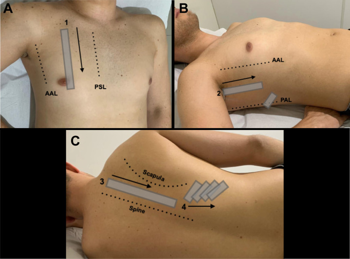
Proposed scanning protocol, placing extra emphasis on the posterior lung regions given the tendency of coronavirus disease 2019 to manifest in these areas. A, Zone 1, the intercostal spaces in the mid-clavicular line. B, Zone 2, the intercostal spaces in the mid-axillary line, plus the area just above the diaphragm in the posterior axillary line. C, Zone 3 (the intercostal spaces between the spine and scapula) and Zone 4 (the area beneath the scapula). AAL = anterior axillary line; PAL = posterior axillary line; PSL = parasternal line.

A-F, Typical lung ultrasound findings in coronavirus disease 2019. A, Multiple B-lines (arrows), seen using a convex transducer oriented parallel to the ribs. B, Multiple B-lines (arrows) and a thickened irregular pleural line (dotted arrow), seen using a convex transducer oriented perpendicular to the ribs. C, Thickened irregular pleura (arrow) and an associated small pleural effusion (dotted arrow), seen using a linear transducer oriented perpendicular to the ribs. D, Thickened irregular pleura (arrow), seen using a phased-array transducer oriented perpendicular to the ribs. E, Multiple B-lines (arrows), seen using a phased-array transducer oriented perpendicular to the ribs. F, A subpleural consolidation (arrow), seen using a phased-array transducer oriented perpendicular to the ribs.
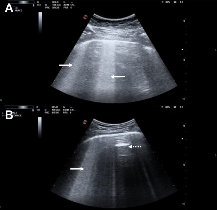
A-B, Two examples of the “light beam” artifact, both generated using the convex ultrasound transducer oriented parallel to the ribs. Areas of thick confluent B-lines can be seen (arrows), emanating from a pleural line that is relatively spared from inflammation, along with interspaced regions of normally aerated lungs where A-lines are seen (spared areas or “skip” lesions; dotted arrows).
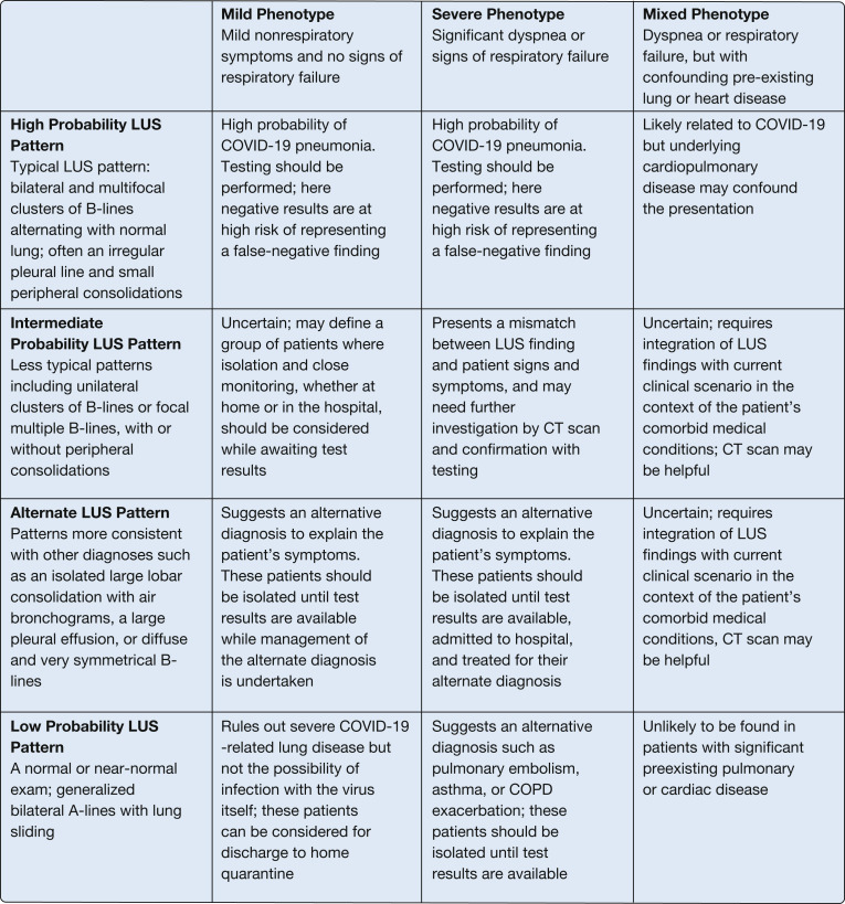
Combining the clinical phenotype with the LUS pattern to aid in patient triage. COVID-19 = coronavirus disease 2019; LUS = lung ultrasound.
References
Publication types
MeSH terms
LinkOut - more resources
Full Text Sources
Other Literature Sources
Medical
