Review
. 2020 Nov:63:101151.
doi: 10.1016/j.arr.2020.101151. Epub 2020 Aug 22.

The ageing kidney: Molecular mechanisms and clinical implications

Affiliations
Review

The ageing kidney: Molecular mechanisms and clinical implications

Yudong Fang et al. Ageing Res Rev. 2020 Nov.

Abstract

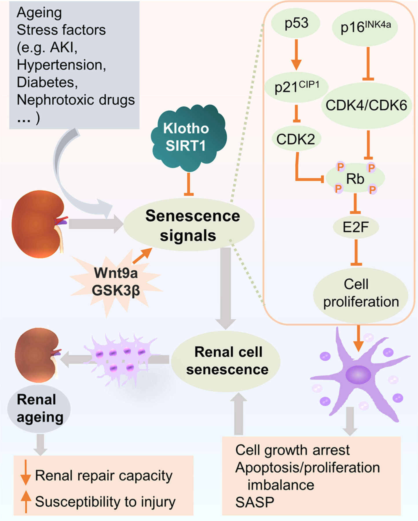
As human life expectancy keeps increasing, ageing populations present a growing challenge for clinical practices. Human ageing is associated with molecular, structural, and functional changes in a variety of organ systems, including the kidney. During the ageing process, the kidney experiences progressive functional decline as well as macroscopic and microscopic histological alterations, which are accentuated by systemic comorbidities like hypertension and diabetes mellitus, or by preexisting or underlying kidney diseases. Although ageing per se does not cause kidney injury, physiologic changes associated with normal ageing processes are likely to impair the reparative capacity of the kidney and thus predispose older people to acute kidney disease, chronic kidney disease and other renal diseases. Mechanistically, cell senescence plays a key role in renal ageing, involving a number of cellular signaling mechanisms, many of which may be harnessed as international targets for slowing or even reversing kidney ageing. This review summarizes the clinical characteristics of renal ageing, highlights the latest progresses in deciphering the role of cell senescence in renal ageing, and envisages potential interventional strategies and novel therapeutic targets for preventing or improving renal ageing in the hope of maintaining long-term kidney health and function across the life course.

Keywords: Glomeruli; Kidney diseases; Kidney transplantation; Nephrosclerosis; Renal tubules; Senescence.


Conflict of interest statement

Declaration of Competing Interest

The authors declare that they have no conflict of interest.

Figures

Fig. 1.
Fig. 1.
Histological characteristics of the ageing kidney. (a) Representative light micrographs showing normal histological characteristics in the healthy young kidney (periodic acid-Schiff staining, ×100). (a1) The enlarged view showing a normal glomerulus. Scale bar, 100 μm. (b) Representative light micrographs showing histological characteristics in the ageing kidney, including ischemic global glomerulosclerosis, focal tubular atrophy and interstitial fibrosis, and arteriolar hyalinosis (periodic acid-Schiff staining, ×100). (b1) Representative light micrograph highlighting nephrosclerosis in the ageing kidney. The arrow indicates global glomerulosclerosis, black arrowheads indicate focal tubular atrophy and interstitial fibrosis, and green arrowheads indicate arteriolar hyalinosis. (b2) The enlarged view showing a moderately hypertrophic glomerulus. Scale bar, 100 μm.
Fig. 2.
Fig. 2.
Cellular signaling pathways involved in cell senescence in renal ageing. In renal ageing, cell senescence signaling pathways are activated not only by stress factors or diseases, such as AKI, hypertension, diabetes, and cytotoxic drugs, etc, but also by ageing per se. The key signaling cascades implicated in renal cell senescence are p53/p21CIP1 and p16INK4a/Rb pathways, which in turn inhibit CDK complexes and Rb phosphorylation. Ultimately, these signals execute cell senescence via Rb suppression of the activity of E2F, characterized by cell proliferation arrest, apoptosis/proliferation imbalance and secretion of SASP factors. Hence, the repair capacity declines and the ageing kidney becomes more susceptible to injury. Klotho and SIRT1 have been shown as main modulators of cell senescence and inhibit cell senescence through regulating p53/p21CIP1 pathway. Whereas, Wnt9a accelerates renal fibrosis via promoting cell senescence signaling pathways, so does GSK3β. Abbreviations: AKI, acute kidney disease; CDK, cyclin-dependent kinase; GSK3β, glycogen synthase kinase 3β; Rb, retinoblastoma protein; SASP, senescence-associated secretory phenotype.
Fig. 3.
Fig. 3.
Categorization of cell senescence. Two main types of cell senescence have been identified according to the different causes, i.e. replicative senescence, and stress-induced premature senescence (SIPS). Replicative senescence is caused by telomere attrition and characterized by low response in proliferation and ultimately cell-cycle arrest. SIPS is induced by various stressors, such as oxidative stress, DNA damage, mitochondrial dysfunction, epigenetic stress, and senescence-associated secretory phenotype (SASP) generated by primary senescent cells. There are also two classes of senescent cells in the process of senescence, i.e. acute and chronic senescent cells, which play different roles in kidney ageing. Acute senescence acts a beneficial role in renal regeneration after injury, renal fibrosis, immune surveillance, and wound healing, where the senescent cells transiently present and eventual eliminated by immune cells through immune surveillance process. In contrast, chronic senescence is elicited due to abnormal accumulation of senescent cells, inefficient clearance, or prolonged senescent signaling. It exerts deleterious effects in natural kidney ageing and age-related kidney diseases.
Fig. 4.
Fig. 4.
Changes of glomerular podocytes in kidney ageing. Transmission electron micrographs show (a) the typical morphology of normal glomerular podocytes in the healthy young kidney. Scale bar, 2 μm. (b) ultrastructural changes of glomerular podocytes in the ageing kidney, characterized by variable foot processes effacement (green arrowhead), podocyte detachment, cytoplasmatic absorption droplets (blue arrowhead), concomitant with thickening of glomerular basement membrane (red arrowhead). Scale bar, 2 μm.

References

    1. Acosta JC, Banito A, Wuestefeld T, Georgilis A, Janich P, Morton JP, Athineos D, Kang TW, Lasitschka F, Andrulis M, Pascual G, Morris KJ, Khan S, Jin H, Dharmalingam G, Snijders AP, Carroll T, Capper D, Pritchard C, Inman GJ, Longerich T, Sansom OJ, Benitah SA, Zender L, Gil J, 2013. A complex secretory program orchestrated by the inflammasome controls paracrine senescence. Nat. Cell Biol. 15, 978–990. - PMC - PubMed
    1. Administration for Community Living, 2019. 2018 profile of older Americans U.S. Department of Health and Human Services.
    1. Al-Douahji M, Brugarolas J, Brown PA, Stehman-Breen CO, Alpers CE, Shankland SJ, 1999. The cyclin kinase inhibitor p21WAF1/CIP1 is required for glomerular hypertrophy in experimental diabetic nephropathy. Kidney Int 56, 1691–1699. - PubMed
    1. Al-Said J, O’Neill WC, 2003. Reduced kidney size in patients with simple renal cysts. Kidney Int 64, 1059–1064. - PubMed
    1. Al-Said J, Brumback MA, Moghazi S, Baumgarten DA, O’Neill WC, 2004. Reduced renal function in patients with simple renal cysts. Kidney Int 65, 2303–2308. - PubMed

Publication types