

A Deep Learning System to Screen Novel Coronavirus Disease 2019 Pneumonia
- PMID: 32837749
- PMCID: PMC7320702
- DOI: 10.1016/j.eng.2020.04.010
A Deep Learning System to Screen Novel Coronavirus Disease 2019 Pneumonia
Abstract
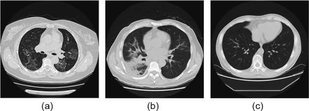
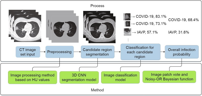
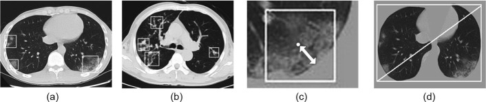
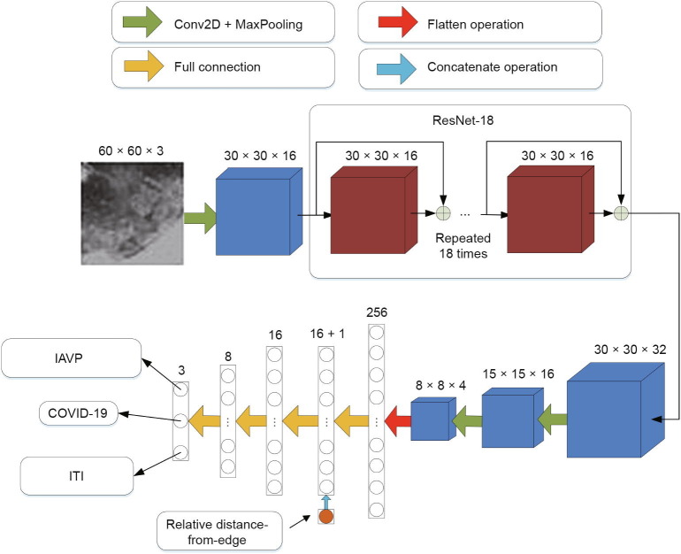
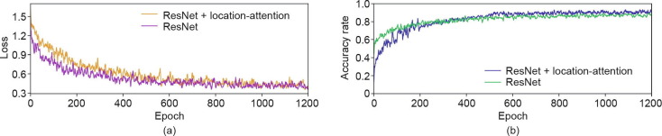
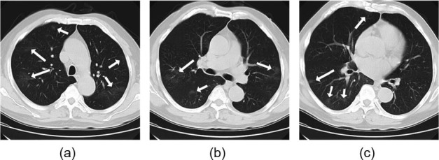
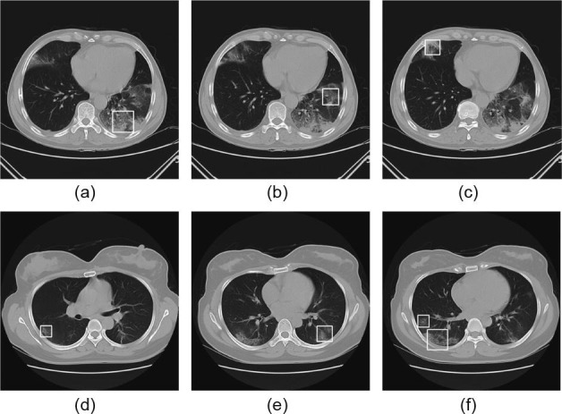
The real-time reverse transcription-polymerase chain reaction (RT-PCR) detection of viral RNA from sputum or nasopharyngeal swab had a relatively low positive rate in the early stage of coronavirus disease 2019 (COVID-19). Meanwhile, the manifestations of COVID-19 as seen through computed tomography (CT) imaging show individual characteristics that differ from those of other types of viral pneumonia such as influenza-A viral pneumonia (IAVP). This study aimed to establish an early screening model to distinguish COVID-19 from IAVP and healthy cases through pulmonary CT images using deep learning techniques. A total of 618 CT samples were collected: 219 samples from 110 patients with COVID-19 (mean age 50 years; 63 (57.3%) male patients); 224 samples from 224 patients with IAVP (mean age 61 years; 156 (69.6%) male patients); and 175 samples from 175 healthy cases (mean age 39 years; 97 (55.4%) male patients). All CT samples were contributed from three COVID-19-designated hospitals in Zhejiang Province, China. First, the candidate infection regions were segmented out from the pulmonary CT image set using a 3D deep learning model. These separated images were then categorized into the COVID-19, IAVP, and irrelevant to infection (ITI) groups, together with the corresponding confidence scores, using a location-attention classification model. Finally, the infection type and overall confidence score for each CT case were calculated using the Noisy-OR Bayesian function. The experimental result of the benchmark dataset showed that the overall accuracy rate was 86.7% in terms of all the CT cases taken together. The deep learning models established in this study were effective for the early screening of COVID-19 patients and were demonstrated to be a promising supplementary diagnostic method for frontline clinical doctors.
Keywords: COVID-19; Computed tomography; Location-attention classification model.
© 2020 THE AUTHORS.
Figures
References
-
- Cohen J., Normile D. New SARS-like virus in China triggers alarm. Science. 2020;367(6475):234–235. - PubMed
LinkOut - more resources
Full Text Sources
Other Literature Sources