. 2020 Dec;7(4):551-557.
doi: 10.1016/j.gendis.2020.07.006. Epub 2020 Jul 17.

Development of cell-based pseudovirus entry assay to identify potential viral entry inhibitors and neutralizing antibodies against SARS-CoV-2

Affiliations

Development of cell-based pseudovirus entry assay to identify potential viral entry inhibitors and neutralizing antibodies against SARS-CoV-2

Jie Hu et al. Genes Dis. 2020 Dec.

Abstract

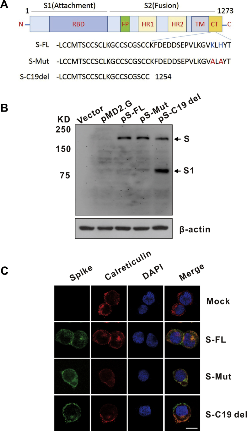
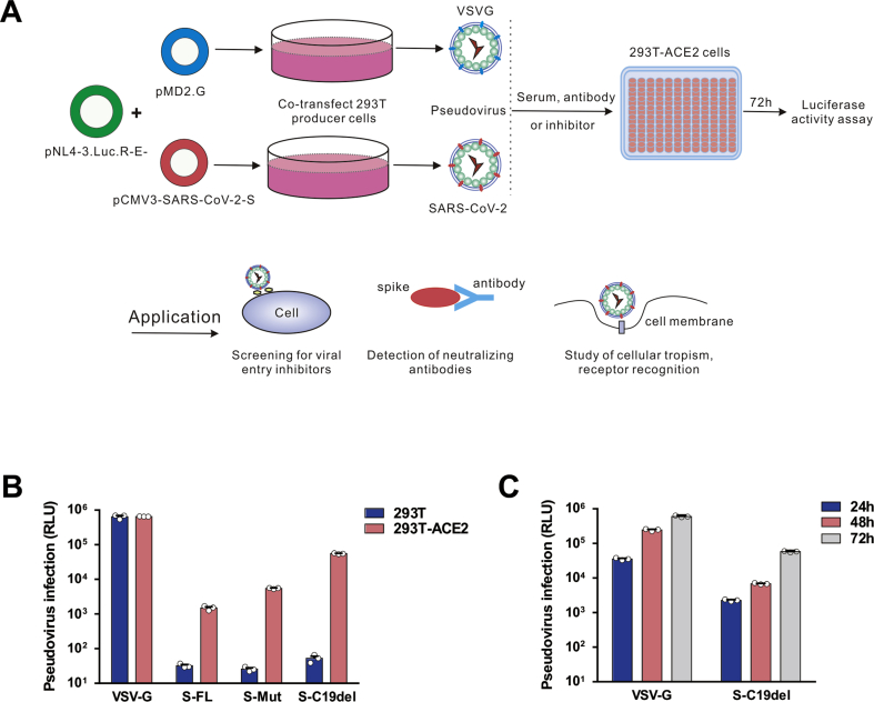
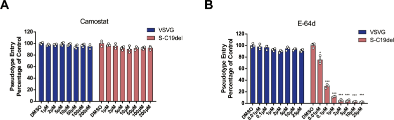
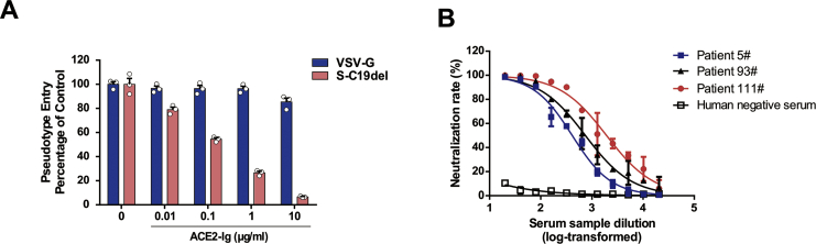
Severe acute respiratory syndrome coronavirus 2 (SARS-CoV-2) is the causative virus of the coronavirus disease 2019 (COVID-19) pandemic. To establish a safe and convenient assay system for studying entry inhibitors and neutralizing antibodies against SARS-CoV-2, we constructed a codon-optimized, full-length C-terminal mutant spike (S) gene of SARS-CoV-2. We generated a luciferase (Luc)-expressing pseudovirus containing the wild-type or mutant S protein of SARS-CoV-2 in the envelope-defective HIV-1 backbone. The key parameters for this pseudovirus-based assay, including the S mutants and virus incubation time, were optimized. This pseudovirus contains a Luc reporter gene that enabled us to easily quantify virus entry into angiotensin-converting enzyme 2 (ACE2)-expressing 293T cells. Cathepsin (Cat)B/L inhibitor E-64d could significantly block SARS-CoV-2 pseudovirus infection in 293T-ACE2 cells. Furthermore, the SARS-CoV-2 spike pseudotyped virus could be neutralized by sera from convalescent COVID-19 patients or recombinant ACE2 with the fused Fc region of human IgG1. Thus, we developed a pseudovirus-based assay for SARS-CoV-2, which will be valuable for evaluating viral entry inhibitors and neutralizing antibodies against this highly pathogenic virus.

Keywords: Antiviral therapeutics; Coronavirus; Neutralizing antibodies; Pseudovirus; SARS-CoV-2; Spike protein.


Conflict of interest statement

The authors declare no competing interests.

Figures

Figure 1
Figure 1
Detection of SARS-CoV-2 spike (S) protein expression and localization. (A) Schematic illustration of the SARS-CoV-2 full-length spike (S-FL) and mutant S variants. The RBD (receptor binding domain) is in subunit S1; the FP (fusion peptide), HR1 (heptad repeat 1), HR2 (heptad repeat 2), TM (transmembrane domain), and CT (cytoplasmic tail) are in subunit S2. The endoplasmic reticulum retrieval signals (“KxHxx” motif) in the CT domain of S-FL were destroyed in S-Mut protein. The C-terminal 19 amino acids were lacking in S-C19del. (B) Detection of SARS-CoV-2 S expression in HKE293T cells by Western blot using the anti-RBD monoclonal antibody. Cells were transfected with pS-FL, pS-Mut, and pS-C19del plasmids or with an empty vector. (C) Detection of SARS-CoV-2 S subcellular localization in HKE293T cells by confocal microscopy. Cells were grown on glass coverslips for 24 h preceding transfection of plasmids encoding S protein variants. The cells were harvested and labeled with the corresponding antibodies. Calreticulin, ER marker. Nuclei were counterstained with DAPI. Bar = 20 μm.
Figure 2
Figure 2
Detection of SARS-CoV-2 S pseudotyped virus infectivity. (A) Schematic representation of the pseudovirus production and neutralization assay and the applications of the pseudovirus. (B) HEK293T and 293T-ACE2 cells were infected with lentiviruses pseudotyped with vesicular stomatitis virus G (VSV-G) and SARS-CoV-2 S protein variants. The y-axis shows the relative luminescence units (RLU) detected at 48 h post-pseudovirus inoculation. (C) Optimization of the incubation time for pseudovirus luciferase assay. Luciferase activities were measured 24–72 h post-virus infection. For this purpose, 72 h was chosen as the optimized incubation time. The data are presented as the means ± standard deviations (SDs) of three independent biological replicates.
Figure 3
Figure 3
Detection of entry inhibitors against SARS-CoV-2 pseudovirus infection. (A–B) Inhibitors camostat mesylate and E-64d blocked SARS-CoV-2 pseudovirus entry. 293T-ACE2 cells were pre-incubated with camostat mesylate (A) or E-64d (B), and subsequently inoculated with pseudovirions. The VSV-G pseudovirus was used as the control. RLU were detected at 72 h post-pseudovirus inoculation. Cell viability was examined by the methyl thiazolyl tetrazolium (MTT) assay. The data are presented as the means ± SDs of three independent biological replicates.
Figure 4
Figure 4
Detection of neutralizing antibodies in sera against SARS-CoV-2 pseudovirus infection. (A) Inhibition of SARS-CoV-2 pseudovirus entry by ACE2-Ig. S-C19del or VSV-G pseudovirions were preincubated with ACE2-Ig and added to 293T-ACE2 cells. (B) Sera from convalescent COVID-19 patients neutralized the SARS-CoV-2 pseudovirus. Serum sample from healthy individual was tested as a negative control. RLU were detected at 72 h post-pseudovirus inoculation. The data are presented as the mean percentages of inhibition ± SDs of duplicate wells.

References

    1. Masters P.S. Coronaviruses: an overview of their replication and pathogenesis. Adv Virus Res. 2006;66:193–292. - PMC - PubMed
    1. Coronaviridae Study Group of the International Committee on Taxonomy of Viruses The species severe acute respiratory syndrome-related coronavirus: classifying 2019-nCoV and naming it SARS-CoV-2. Nat Microbiol. 2020;5:536–544. - PMC - PubMed
    1. Zhou P., Yang X.-L., Wang X.-G. A pneumonia outbreak associated with a new coronavirus of probable bat origin. Nature. 2020;579:270–273. - PMC - PubMed
    1. Wrapp D., Wang N., Corbett K.S. Cryo-EM structure of the 2019-nCoV spike in the prefusion conformation. Science. 2020;367:1260–1263. - PMC - PubMed
    1. Walls A.C., Park Y.-J., Tortorici M.A., Wall A., McGuire A.T., Veesler D. Structure, function, and antigenicity of the SARS-CoV-2 spike glycoprotein. Cell. 2020;181 281-292.e6. - PMC - PubMed