. 2020:20:100413.
doi: 10.1016/j.imu.2020.100413. Epub 2020 Aug 13.

SARS-CoV-2-human protein-protein interaction network

Affiliations

SARS-CoV-2-human protein-protein interaction network

Babak Khorsand et al. Inform Med Unlocked. 2020.

Abstract

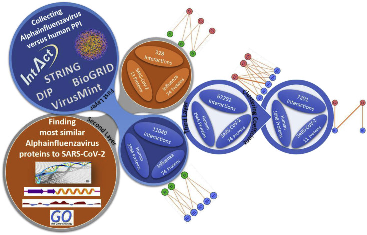
Severe acute respiratory syndrome coronavirus 2 (SARS-CoV-2) is the novel coronavirus which caused the coronavirus disease 2019 pandemic and infected more than 12 million victims and resulted in over 560,000 deaths in 213 countries around the world. Having no symptoms in the first week of infection increases the rate of spreading the virus. The increasing rate of the number of infected individuals and its high mortality necessitates an immediate development of proper diagnostic methods and effective treatments. SARS-CoV-2, similar to other viruses, needs to interact with the host proteins to reach the host cells and replicate its genome. Consequently, virus-host protein-protein interaction (PPI) identification could be useful in predicting the behavior of the virus and the design of antiviral drugs. Identification of virus-host PPIs using experimental approaches are very time consuming and expensive. Computational approaches could be acceptable alternatives for many preliminary investigations. In this study, we developed a new method to predict SARS-CoV-2-human PPIs. Our model is a three-layer network in which the first layer contains the most similar Alphainfluenzavirus proteins to SARS-CoV-2 proteins. The second layer contains protein-protein interactions between Alphainfluenzavirus proteins and human proteins. The last layer reveals protein-protein interactions between SARS-CoV-2 proteins and human proteins by using the clustering coefficient network property on the first two layers. To further analyze the results of our prediction network, we investigated human proteins targeted by SARS-CoV-2 proteins and reported the most central human proteins in human PPI network. Moreover, differentially expressed genes of previous researches were investigated and PPIs of SARS-CoV-2-human network, the human proteins of which were related to upregulated genes, were reported.

Keywords: COVID-19; Coronavirus; Host-pathogen protein interaction; Protein interaction prediction; Protein-protein interaction; SARS-CoV-2.


Conflict of interest statement

The authors declare that they have no known competing financial interests or personal relationships that could have appeared to influence the work reported in this paper.

Figures

Fig. 1
Fig. 1
Fig. 1-a shows the spreading speed of COVID-19 confirmed cases of different countries in reaching 300000 cases, while 1-c shows the time that each country needs to sacrifice 30000 cases by COVID-19. Fig. 1-b shows the total COVID-19 confirmed cases of different countries, and 1-d shows the total number of deaths caused by COVID-19.
Fig. 2
Fig. 2
Fig. 2-a shows the number of deaths from each 1000 COVID-19 cases (death rate). Fig. 2-b shows the number of COVID-19 cases per each 100000 individuals of the respective countries.
Fig. 3
Fig. 3
Fig. 3-a shows the whole spreading distribution of COVID-19. Fig. 3-b and 3-c shows the daily spreading and death distributions, respectively.
Fig. 4
Fig. 4
Schematic view of predicting SARS-CoV-2-human protein-protein interaction network.
Fig. 5
Fig. 5
A multipartite network in which the first layer shows the similarity between AIV proteins (I1, I2, and I3) and SV proteins (C1, C2, and C3) with green edges. The second layer shows the interactions between AIV proteins and human proteins (H1, H2, H3, H4, and H5) with blue edges. The third layer shows the possible interactions between SV proteins and human proteins with red edges. The thicknesses of the three red edge types shows that the ticker ones could be better candidates for SV-human PPI network. (For interpretation of the references to color in this figure legend, the reader is referred to the Web version of this article.)
Fig. 6
Fig. 6
Fig. 6-a shows the amino acid composition of SARS-CoV-2 proteins. Fig. 6-b shows the whole amino acid distribution of SARS-CoV-2 proteins.
Fig. 7
Fig. 7
Fig. 7-a shows the secondary structure of SARS-CoV-2 proteins. Light, medium, and dark pink declare coil, helix, and extended structures, respectively. Fig. 7-b shows the accessible surface area of SARS-CoV-2 proteins. Two lowest colors of gradient represent buried residues and the other colors of gradient show exposed residues. (For interpretation of the references to color in this figure legend, the reader is referred to the Web version of this article.)
Fig. 8
Fig. 8
Fig. 8-a shows P0DTC7-human protein-protein interaction network. P0DTC7 proteins are shown with red nodes. Its IAV orthologs are shown with green nodes which have 389 interactions with 218 HPs (blue nodes). 49 HPs which have weights more than 10 and are connected to at least 2 IAV proteins were selected as final interactors of P0DTC7 proteins and so the final P0DTC7-human PPI network is a bipartite network among the red nodes and 49 blue nodes which are connected by red edges. Fig. 8-b shows induced subgraph of human protein-protein interaction network of human proteins interacting with SARS-CoV-2 proteins. (For interpretation of the references to color in this figure legend, the reader is referred to the Web version of this article.)
Fig. 9
Fig. 9
122 enriched molecular functions of human proteins targeted by SARS-CoV-2 proteins.

References

    1. Khani H., Tabarraei A., Moradi A. Survey of coronaviruses infection among patients with flu-like symptoms in the golestan province, Iran TT - mljgoums. Nov. 2018;12(6):1–4.
    1. Qu J., Wickramasinghe C. SARS, MERS and the sunspot cycle. Curr Sci. 2017;113(8):1501.
    1. Luk H.K.H., Li X., Fung J., Lau S.K.P., Woo P.C.Y. Molecular epidemiology, evolution and phylogeny of SARS coronavirus. Infect Genet Evol. 2019;71:21–30. - PMC - PubMed
    1. Organization W.H. 2003. Summary of probable SARS cases with onset of illness from 1 November 2002 to 31 July 2003.
    1. Dudas G., Carvalho L.M., Rambaut A., Bedford T. MERS-CoV spillover at the camel-human interface. Elife. 2018;7 - PMC - PubMed

LinkOut - more resources