

Case Reports
doi: 10.1016/j.ekir.2020.07.008.
Epub 2020 Jul 16.
Oxalate Nephropathy Caused by Excessive Vitamin C Administration in 2 Patients With COVID-19
Affiliations
- PMID: 32838081
- PMCID: PMC7363608
- DOI: 10.1016/j.ekir.2020.07.008
Item in Clipboard
Case Reports
Oxalate Nephropathy Caused by Excessive Vitamin C Administration in 2 Patients With COVID-19
Kidney Int Rep.
2020 Oct.
No abstract available
Figures
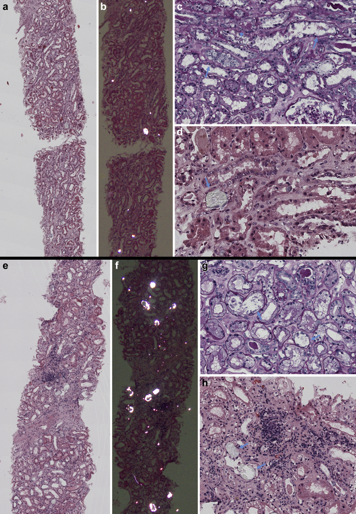
Light microscopy examination of kidney biopsy specimens obtained from patients 1 (a–d) and 2 (e–h). (a,e) Low-power view of kidney biopsy specimens with focus on tubule interstitium; several crystals are in the tubular lumens (hematoxylin and eosin [H&E] stain). (b,f) Same sections as a and c examined under polarized light: several birefringent crystals are visible in the tubular lumina (H&E stain). (c) Diffuse acute tubular injury with flattening of epithelial cell line (arrows) and focal cell detachment with intraluminal sloughing (arrowhead) (periodic acid–Schiff stain; original magnification ×400). (d) Detail of intratubular calcium oxalate crystal (arrow), with translucent appearance (H&E stain; original magnification ×400). (g) Diffuse acute tubular injury with extensive vacuolization of tubular epithelial cells and focal flattening of epithelial line (arrows) (periodic acid–Schiff stain; original magnification ×400). (h) Detail of intratubular calcium oxalate crystal (arrow), with translucent appearance (arrows); moderate mixed interstitial infiltrate in area of initial interstitial fibrosis (H&E stain; original magnification ×400).
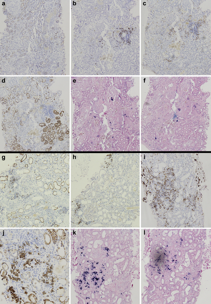
Immunohistochemistry characterization of interstitial inflammatory infiltrate of patients 1 (a–f) and 2 (g–l). Patient 1 showed minimal scattered interstitial infiltrate, composed mainly of B lymphocytes (b) and macrophages (c); patient 2 showed focal moderate interstitial inflammation with abundant presence of plasma cells (j) and macrophages (i); both T (g) and B (h) lymphocytes were present. (a,g) CD3 (T-lymphocytes; original magnification ×100); (b,h) CD20 (B-lymphocytes; original magnification ×100); (c,i) CD163 (macrophages; original magnification ×100); (d,j) CD138 (plasma cells; original magnifications: d, ×100; j, ×200); (e,k) kappa light chain (original magnification ×100); and (f,l) lambda light chain (original magnification ×100).
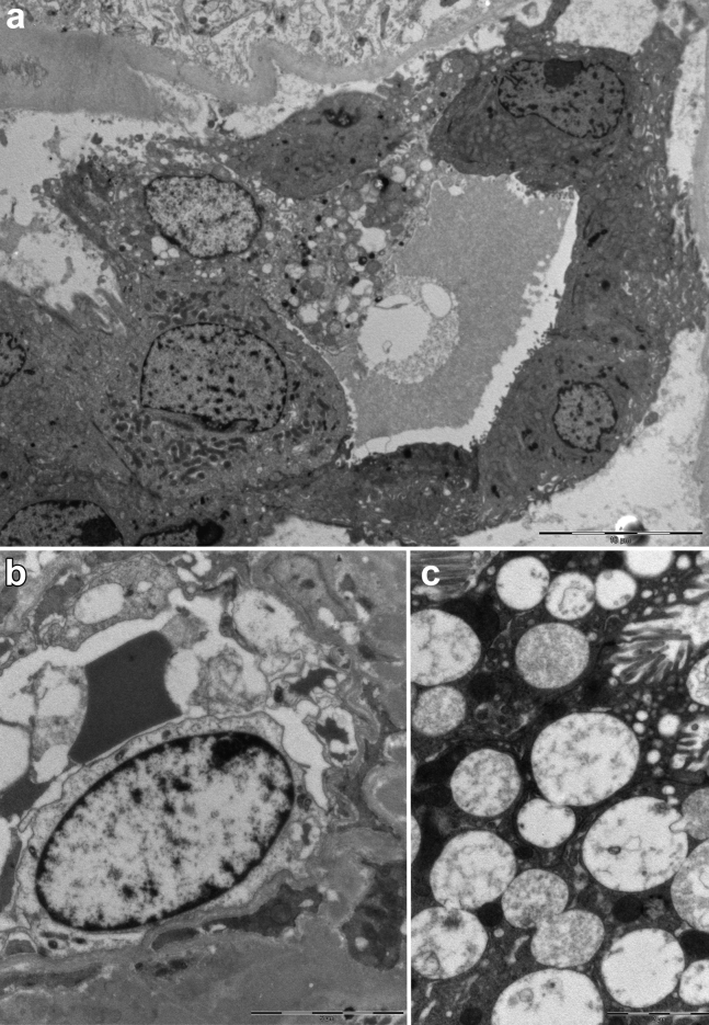
Ultrastructural analysis on transmission electron microscopy of kidney biopsy of patient 2. (a) Low magnification of a distal tubule with diffuse signs of acute injury (detachment of cells from tubular basement membrane; bar = 10 μm). (b) Peritubular capillary structure lined by a degenerated necrotic endothelial cell (bar = 5 μm). (c) Cytoplasmic vacuoles in a tubular epithelial cell; vacuoles are polymorphic in appearance, most often with low electron density content comprised of lamellar and corpuscular figures that are not suggestive for viral structures (diameter, >130 nm; bar = 2 μm).
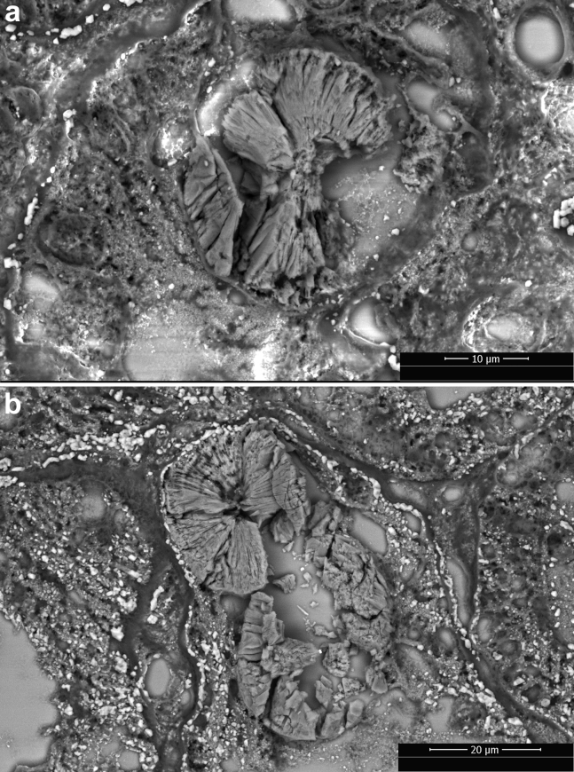
Scanning electron microscope photographs of calcium oxalate monohydrate crystals in the tubular lumen in the kidney biopsy specimen obtained from patients 1 (a) and 2 (b). Crystals show the typical rose-cut–shaped appearance.
References
Publication types
LinkOut - more resources
Full Text Sources
