. 2020 Sep 23;12(38):42499-42510.
doi: 10.1021/acsami.0c10781. Epub 2020 Sep 8.

Polydopamine-Mesoporous Silica Core-Shell Nanoparticles for Combined Photothermal Immunotherapy

Affiliations

Polydopamine-Mesoporous Silica Core-Shell Nanoparticles for Combined Photothermal Immunotherapy

Anushree Seth et al. ACS Appl Mater Interfaces. .

Abstract

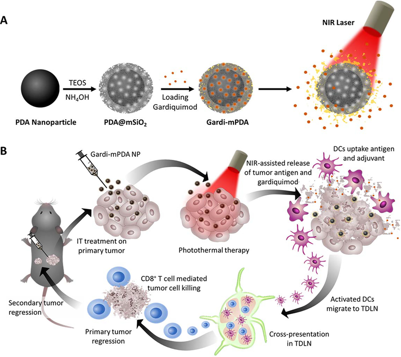
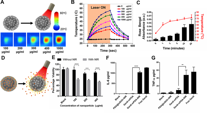
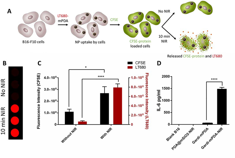
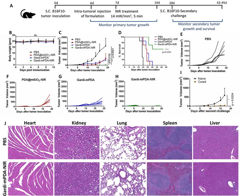
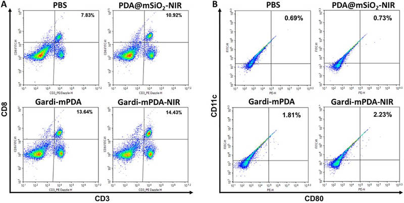
Cancer immunotherapy involves a cascade of events that ultimately leads to cytotoxic immune cells effectively identifying and destroying cancer cells. Responsive nanomaterials, which enable spatiotemporal orchestration of various immunological events for mounting a highly potent and long-lasting antitumor immune response, are an attractive platform to overcome challenges associated with existing cancer immunotherapies. Here, we report a multifunctional near-infrared (NIR)-responsive core-shell nanoparticle, which enables (i) photothermal ablation of cancer cells for generating tumor-associated antigen (TAA) and (ii) triggered release of an immunomodulatory drug (gardiquimod) for starting a series of immunological events. The core of these nanostructures is composed of a polydopamine nanoparticle, which serves as a photothermal agent, and the shell is made of mesoporous silica, which serves as a drug carrier. We employed a phase-change material as a gatekeeper to achieve concurrent release of both TAA and adjuvant, thus efficiently activating the antigen-presenting cells. Photothermal immunotherapy enabled by these nanostructures resulted in regression of primary tumor and significantly improved inhibition of secondary tumor in a mouse melanoma model. These biocompatible, biodegradable, and NIR-responsive core-shell nanostructures simultaneously deliver payload and cause photothermal ablation of the cancer cells. Our results demonstrate potential of responsive nanomaterials in generating highly synergistic photothermal immunotherapeutic response.

Keywords: NIR-responsive drug delivery; cancer immunotherapy; mesoporous silica; photothermal therapy; polydopamine nanoparticles.


Conflict of interest statement

The authors declare no competing financial interests.

Supporting Information

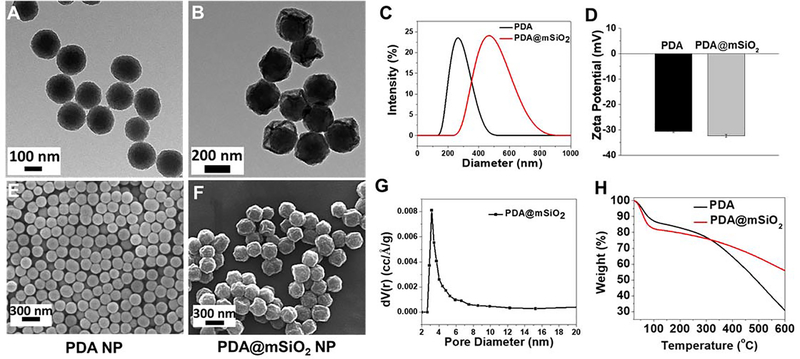
Analysis of TGA data; Loading and release of LT680; Photothermal efficiency calculation; SEM image of as-prepared silica-coated PDA nanoparticles before removing the pore-template (CTAB); HRTEM image of PDA@mSiO2 nanoparticles and corresponding EDS elemental mapping for N and Si; Absorbance spectra of PDA and PDA@mSiO2 nanoparticles; TEM images of the PDA@mSiO2 nanoparticles before and after 10 minutes laser treatment; In vitro maturation of BMDCs; Fluorescence images showing efficient dye loading without free dye in solution and NIR-triggered release of LT680 dye from PDA@mSiO2; Nanoparticle uptake by cancer cells; Absorption and emission spectra of CFSE and LT680; Infrared images, fluorescence images and changes in local temperature of the mouse melanoma model; Relative populations and activation status of immune cells in tumor draining lymph node; Therapeutic effect of increasing NIR treatment duration to 10 minutes.

Figures

Figure 1.
Figure 1.
Schematic illustrations depicting (A) Synthesis of gardiquimod-loaded mesoporous silica coated polydopamine nanoparticles (gardi-mPDA) and NIR-assisted drug release. (B) Tumor ablation and drug release under NIR irradiation followed by activation of DCs and effector T cells in tumor draining lymph nodes for regression of primary and secondary tumors.
Figure 2.
Figure 2.
TEM images of the (A) PDA and (B) PDA@mSiO2 nanoparticles. (C) hydrodynamic diameter and (D) zeta potential of the PDA and PDA@mSiO2 nanoparticles. SEM images of the (E) PDA and (F) PDA@mSiO2 nanoparticles. (G) Pore size distribution of PDA@mSiO2 nanoparticles obtained by nitrogen adsorption and using Barrett-Joyner-Halenda (BJH) method. (H) Weight loss profiles of PDA and PDA@mSiO2 nanoparticles as measured by thermogravimetric analysis.
Figure 3.
Figure 3.
(A) Schematic representation of NIR irradiation of pristine PDA@mSiO2 nanoparticles and IR images of temperature rise with increase in particle concentration after 5 minutes of NIR laser treatment. (B) Temperature profile and effect of PDA@mSiO2 particle concentration on temperature rise when aqueous solutions were subjected to laser power density of 14 mW/mm2. (C) Cumulative release of model dye from the PDA@mSiO2 nanoparticles after different laser irradiation durations and their corresponding solution temperature (laser power density, 14 mW/mm2). (D) Schematic representation of gardiquimod loaded PDA@mSiO2 (gardi-mPDA) nanoparticles and release of cargo with NIR treatment. (E) Cancer cell viability after treatment with gardi-mPDA with and without NIR. BMDC activation indicated by cytokine secretion (F) IL-6 and (G) TNFα. Data represented as mean ± SD. ** p<0.01, *** p<0.001 and **** p<0.0001 by one-way ANOVA with Tukey’s posttest.
Figure 4.
Figure 4.
Combined photothermal-immunotherapy effect in the presence of NIR. (A) Schematic illustration describing the experiment. Briefly, B16-F10 cells were treated with LT680-mPDA followed by labelling with CFSE. The labelled cells were divided into 2 groups and one group was given NIR treatment for 10 min. The supernatant of 2 groups were collected after 12 hours and fluorescence intensity was measured. (B) Fluorescence images (LT680) of supernatants collected from cells treatment with and without NIR, (C) Fluorescence intensity of CFSE and LT680 with and without NIR, (D) IL-6 secretion by BMDCs treated with supernatants released from B16-F10 cells. Data represented as mean ± SD. * p<0.05 and **** p<0.0001 by one-way ANOVA with Tukey’s posttest.
Figure 5.
Figure 5.
In vivo photothermal-immunotherapeutic effect of gardi-mPDA.(A) Timeline of experiment. (B) In vivo toxicity of gardi-mPDA and NIR assessed by change in body weight of the mice, (C) tumor growth profiles, (D) survival curve of mice given different treatments (n=7). Tumor growth curves of individual mouse after treatment with (E) PBS, (F) PDA@mSiO2-NIR, (G) gardi-mPDA and (H) gardi-mPDA-NIR. (I) Tumor volume after secondary challenge in mice surviving after gardi-mPDA-NIR treatment (cured mice) and age matched naïve mice (n=3). (J) Hematoxylin−eosin (H&E) staining images of major mice organs after treatment with PBS and gardi-mPDA-NIR. Data represented as mean ± SD. * p<0.05 and **** p<0.0001 by one-way ANOVA with Tukey’s posttest and Log-rank (Mantel-Cox) test for survival curve.
Figure 6.
Figure 6.
Relative populations and activation status of immune cells in tumor draining lymph node at day 16. Representative flow cytometry plots of (A) CD3+ CD8+ T cells, (B) CD11c+ CD80+ dendritic cells. Percentage positive cells are displayed on top right corner.

Similar articles

Cited by

References

    1. Vesely MD; Kershaw MH; Schreiber RD; Smyth MJ, Natural Innate and Adaptive Immunity to Cancer. Annu. Rev. Immunol 2011, 29 (1), 235–271. - PubMed
    1. Dunn GP; Bruce AT; Ikeda H; Old LJ; Schreiber RD, Cancer Immunoediting: From Immunosurveillance to Tumor Escape. Nat. Immunol 2002, 3 (11), 991–998. - PubMed
    1. O’Donnell JS; Teng MWL; Smyth MJ, Cancer Immunoediting and Resistance to T Cell-Based Immunotherapy. Nat. Rev. Clin. Oncol 2018. - PubMed
    1. Beatty GL; Gladney WL, Immune Escape Mechanisms as a Guide for Cancer Immunotherapy. Clin. Cancer Res 2015, 21 (4), 687. - PMC - PubMed
    1. Mellman I; Coukos G; Dranoff G, Cancer Immunotherapy Comes of Age. Nature 2011, 480 (7378), 480–489. - PMC - PubMed

MeSH terms