. 2020 Sep:548:39-48.
doi: 10.1016/j.virol.2020.05.015. Epub 2020 Jun 13.

Growth, detection, quantification, and inactivation of SARS-CoV-2

Affiliations

Growth, detection, quantification, and inactivation of SARS-CoV-2

James Brett Case et al. Virology. 2020 Sep.

Abstract

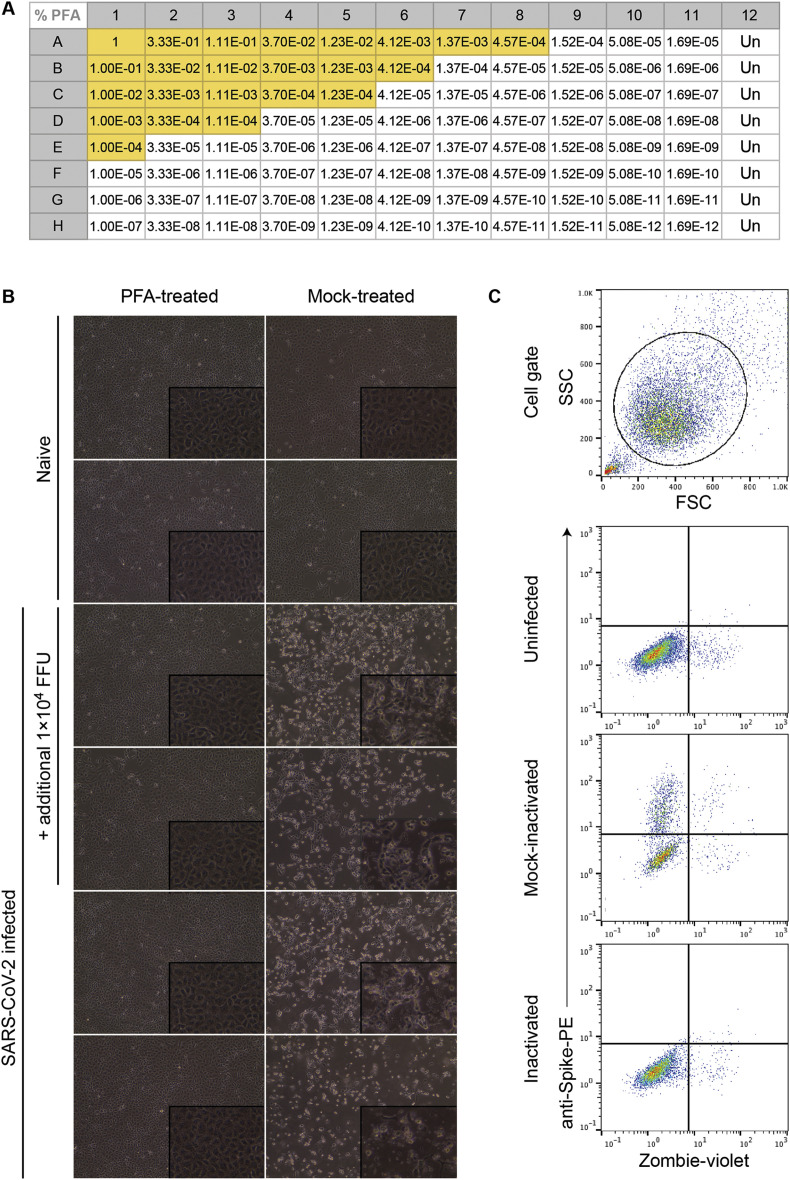
Severe acute respiratory syndrome coronavirus (SARS-CoV)-2 is the agent responsible for the coronavirus disease 2019 (COVID-19) global pandemic. SARS-CoV-2 is closely related to SARS-CoV, which caused the 2003 SARS outbreak. Although numerous reagents were developed to study SARS-CoV infections, few have been applicable to evaluating SARS-CoV-2 infection and immunity. Current limitations in studying SARS-CoV-2 include few validated assays with fully replication-competent wild-type virus. We have developed protocols to propagate, quantify, and work with infectious SARS-CoV-2. Here, we describe: (1) virus stock generation, (2) RT-qPCR quantification of SARS-CoV-2 RNA; (3) detection of SARS-CoV-2 antigen by flow cytometry, (4) quantification of infectious SARS-CoV-2 by focus-forming and plaque assays; and (5) validated protocols for virus inactivation. Collectively, these methods can be adapted to a variety of experimental designs, which should accelerate our understanding of SARS-CoV-2 biology and the development of effective countermeasures against COVID-19.

Keywords: Coronavirus; Flow cytometry; Focus-forming assay; Plaque assay; SARS-CoV-2; Titration; Virus inactivation.


Conflict of interest statement

M.S.D. is a consultant for Inbios, Vir Biotechnology, NGM Biopharmaceuticals, and on the Scientific Advisory Board of Moderna.

Figures

Fig. 1
Fig. 1
SARS-CoV-2 causes cytopathic effect on Vero E6 cell monolayers. Vero E6 cells were inoculated with SARS-CoV-2 at an multiplicity of infection (MOI) of 0.01 plaque forming unit (PFU)/cell and monitored for cytopathic effect at the indicated timepoints. Images were collected using an EVOS XL Core Imaging System. Magnification is 10X for all images.
Fig. 2
Fig. 2
Crystal violet stained plaque assay plates. Vero-furin or Vero E6 cells were inoculated with 10-fold serial dilutions of a SARS-CoV-2 stock. Plates were fixed three days post-infection and stained with crystal violet. Wells with individual plaques were used to determine the virus titer (Vero-furin 10−4, Vero E6 10−3). (For interpretation of the references to color in this figure legend, the reader is referred to the Web version of this article.)
Fig. 3
Fig. 3
SARS-CoV-2 focus-forming assay. CCL81, Vero-furin, Vero E6, and MA104 cells were inoculated with 10-fold serial dilutions of a SARS-CoV-2 stock. Plates were fixed 30 h post-infection and stained with CR3022 anti-SARS-CoV-2 antibody (1 μg/mL) overnight followed by anti-human IgG-HRP (1:500) for 2 h. Foci were visualized using TrueBlue substrate and wells with discrete foci were used to determine virus titer (10−3 - 10−4).
Fig. 4
Fig. 4
SARS-CoV-2 infected cell flow cytometry plots. Indicated cell types were inoculated with SARS-CoV-2 at an MOI of 0.01 PFU/cell. At each indicated timepoint post-infection, cells were collected and prepared for flow cytometry using CR3022 anti-S as the primary antibody followed by goat-anti-human IgG Alexa 647 as the secondary antibody.
Fig. 5
Fig. 5
Virus outgrowth assay. (A) Titration of paraformaldehyde toxicity on Vero E6 cells plated in 96-well format as described in the protocol. Yellow shading indicates wells in which cytopathic effect was observed. (B) Cytopathic effect observed in Vero E6 cells following inoculation with SARS-CoV-2 infected lung homogenate, treated with or without 1% PFA for 60 min and diluted 1:15,000. Photographs show cells under phase-contrast at 20X (and 40X, inset) magnification. (C) Flow cytometric analysis of Vero E6 cells inoculated with SARS-CoV-2 following treatment with an inactivation agent or PBS (mock). Cells were dissociated to single-cell suspension once the mock-treated culture displayed CPE consistent with SARS-CoV-2 infection. Viability staining with Zombie violet was performed prior to fixation. Antibody staining was performed on 4% paraformaldehyde-fixed and permeabilized cells using the CR3022 anti-SARS-CoV-2 spike antibody followed by anti-human IgG-BV421 labelled secondary antibody. (For interpretation of the references to color in this figure legend, the reader is referred to the Web version of this article.)

References

    1. (CDC), C.f.D.C.a.P . 2020. 2019-Novel Coronavirus (2019-nCoV) Real-Time rRT-PCR Panel Primers and Probes.
    1. Corman V.M., Landt O., Kaiser M., Molenkamp R., Meijer A., Chu D.K., Bleicker T., Brunink S., Schneider J., Schmidt M.L., Mulders D.G., Haagmans B.L., van der Veer B., van den Brink S., Wijsman L., Goderski G., Romette J.L., Ellis J., Zambon M., Peiris M., Goossens H., Reusken C., Koopmans M.P., Drosten C. Detection of 2019 novel coronavirus (2019-nCoV) by real-time RT-PCR. Euro Surveill. 2020;25 - PMC - PubMed
    1. Joyce M.G., Sankhala R.S., Chen W.-H., Choe M., Bai H., Hajduczki A., Yan L., Sterling S.L., Peterson C.E., Green E.C., Smith C., de Val N., Amare M., Scott P., Laing E.D., Broder C.C., Rolland M., Michael N.L., Modjarrad K. 2020. A Cryptic Site of Vulnerability on the Receptor Binding Domain of the SARS-CoV-2 Spike Glycoprotein. bioRxiv.
    1. Lei C., Fu W., Qian K., Li T., Zhang S., Ding M., Hu S. 2020. Potent Neutralization of 2019 Novel Coronavirus by Recombinant ACE2-Ig. bioRxiv. - PMC - PubMed
    1. Letko M., Marzi A., Munster V. Functional assessment of cell entry and receptor usage for SARS-CoV-2 and other lineage B betacoronaviruses. Nat. Microbiol. 2020 - PMC - PubMed

Publication types

MeSH terms