

Review
doi: 10.1067/j.cpradiol.2020.06.010.
Epub 2020 Jun 27.
Coronavirus Disease 2019 (COVID-19) Pneumonia Presentations in Chest Computed Tomography: A Pictorial Review
Affiliations
- PMID: 32839069
- PMCID: PMC7320875
- DOI: 10.1067/j.cpradiol.2020.06.010
Item in Clipboard
Review
Coronavirus Disease 2019 (COVID-19) Pneumonia Presentations in Chest Computed Tomography: A Pictorial Review
Curr Probl Diagn Radiol.
2021 May-Jun.
Abstract
Despite imaging not being a tool for novel coronavirus disease 2019 (COVID-19) diagnosis, there has been an increased number of chest computed tomography (CT) scans done worldwide. There are no pathognomonic CT features for COVID-19 pneumonia, as findings are also common in other infectious diseases and noninfectious aetiologies. Nonetheless, point-of-care physicians should be familiarized with the most common imaging presentations of the COVID-19. In this pictorial review, we have summarized the most reported imaging features of COVID-19 pneumonia, including possible differential diagnosis according to the CT finding.
Copyright © 2020. Published by Elsevier Inc.
Figures
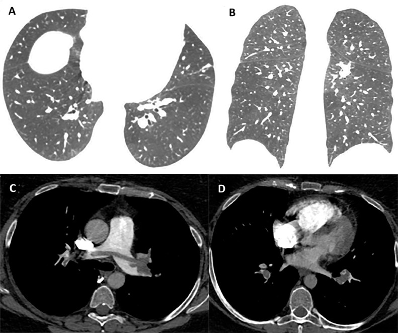
A 38-year-old woman with the diagnosis of COVID-19 by RT-PCR presenting fever for 2 weeks and intense dyspnea for 2 days. (A) axial and (B) coronal CT scans show minimal peripheral GGO in lower lobes. A computed tomography pulmonary angiography (CTPA) was made and showed thrombus in the right and left main pulmonary arteries (C) and in bilateral lower lobe pulmonary arteries (D).

A 49-year-old man with the diagnosis of COVID-19 by RT-PCR presenting fever and diarrhoea for 3 days. (A and B) axial CT scans show a GGO with peripheral and bilateral distribution in lower lobes. (C) Sagittal CT images demonstrated the same findings in the lower lobes.
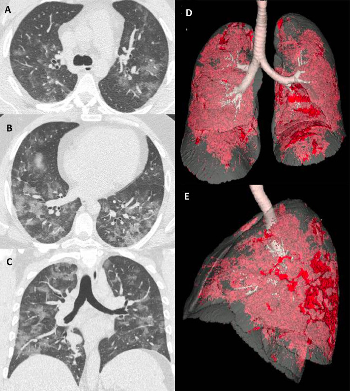
A 63-year-old man with the diagnosis of COVID-19 by RT-PCR presenting fever and cough for 7 days. (A and B) axial and (C) coronal CT scans show a patchy GGO with random distribution in both lungs. (D and E) demonstrate a frontal and lateral view of 3DCT reconstructions with automatic detection of GGO (red). (Color version of figure is available online.)

A 42-year-old woman with the diagnosis of COVID-19 by RT-PCR with dry cough for 8 days. (A) axial and (B) coronal CT scans present rounded GGO in both lungs.

A 34-year-old man with the diagnosis of COVID-19 by RT-PCR presenting fever and diarrhoea for 2 days. The axial CT scan shows a unilateral GGO with peripheral distribution in the right lower lobe.

A 71-year-old woman with the diagnosis of COVID-19 by RT-PCR presenting fever and dyspnea for 9 days. The axial CT scan shows a diffuse GGO with septal thickening, compatible with “crazy paving.”

A 66-year-old man with the diagnosis of COVID-19 by RT-PCR presenting fever and cough for 11 days. (A and B) axial CT scans show areas of consolidation associated with GGO with diffuse distribution in both lungs. (C and D) coronal and sagittal CT reconstructions demonstrated the same findings in lower lobes.

An 82-year-old man with the diagnosis of COVID-19 by RT-PCR presenting fever and diarrhoea for 13 days. He was intubated in day 6 of disease. (A and C) axial CT scans show diffuse consolidation with anteroposterior gradient, compatible with diffuse alveolar damage. (B) CT reconstruction demonstrated the same findings.

A 42-year-old man with the diagnosis of COVID-19 by RT-PCR presenting fever and diarrhoea for 9 days. Axial CT scan show 2 solid nodules surrounded by a ground-glass halo in the right and other in left lower lobes.

A 55-year-old man with the diagnosis of COVID-19 by RT-PCR presenting fever and diarrhoea for 6 days. (A and B) axial CT scans show 2 reversed halo signs in the posterior basal segment of the right and left lower lobe.

A 50-year-old woman with the diagnosis of COVID-19 by RT-PCR with fever and mild dyspnea for 9 days. (A) axial and (B) coronal chest CT image shows multiple rounded GGO with a peripheral ring of consolidation (reversed halo sign).
Similar articles
-
Thoracic imaging in COVID-19.Cleve Clin J Med. 2020 Jul 31;87(8):469-476. doi: 10.3949/ccjm.87a.ccc032. Cleve Clin J Med. 2020. PMID: 32737043
-
COVID-19 mimics on chest CT: a pictorial review and radiologic guide.Br J Radiol. 2021 Feb 1;94(1118):20200703. doi: 10.1259/bjr.20200703. Epub 2020 Dec 9. Br J Radiol. 2021. PMID: 33296607 Free PMC article.
-
Radiological perspective of COVID-19 pneumonia: The early features and progressive behaviour on high-resolution CT.J Med Imaging Radiat Oncol. 2021 Apr;65(2):208-212. doi: 10.1111/1754-9485.13139. Epub 2021 Jan 24. J Med Imaging Radiat Oncol. 2021. PMID: 33491340 Free PMC article. Review.
-
Chest Computed Tomography findings in patients with corona virus disease 2019 (COVID-19): An initial experience in three centres in Ghana, West Africa.J Med Imaging Radiat Sci. 2020 Dec;51(4):604-609. doi: 10.1016/j.jmir.2020.09.005. Epub 2020 Sep 19. J Med Imaging Radiat Sci. 2020. PMID: 33342483 Free PMC article.
-
Chronic Chest Computed Tomography Findings Following COVID-19 Pneumonia.Semin Ultrasound CT MR. 2024 Aug;45(4):298-308. doi: 10.1053/j.sult.2024.02.008. Epub 2024 May 3. Semin Ultrasound CT MR. 2024. PMID: 38704055 Review.
Cited by
-
COVID-19: a brief update for radiologists.Radiol Bras. 2020 Sep-Oct;53(5):320-328. doi: 10.1590/0100-3984.2020.0074. Radiol Bras. 2020. PMID: 33071376 Free PMC article. Review.
-
The importance of chest CT severity score and lung CT patterns in risk assessment in COVID-19-associated pneumonia: a comparative study.Front Med (Lausanne). 2023 May 17;10:1125530. doi: 10.3389/fmed.2023.1125530. eCollection 2023. Front Med (Lausanne). 2023. PMID: 37265487 Free PMC article.
-
Automated Quantitative Lung CT Improves Prognostication in Non-ICU COVID-19 Patients beyond Conventional Biomarkers of Disease.Diagnostics (Basel). 2021 Nov 16;11(11):2125. doi: 10.3390/diagnostics11112125. Diagnostics (Basel). 2021. PMID: 34829472 Free PMC article.
References
Publication types
MeSH terms
LinkOut - more resources
Full Text Sources
Other Literature Sources
Medical
