Review
. 2020 Aug 21;9(9):1941.
doi: 10.3390/cells9091941.

In Vivo TSPO Signal and Neuroinflammation in Alzheimer's Disease

Affiliations
Review

In Vivo TSPO Signal and Neuroinflammation in Alzheimer's Disease

Benjamin B Tournier et al. Cells. .

Abstract

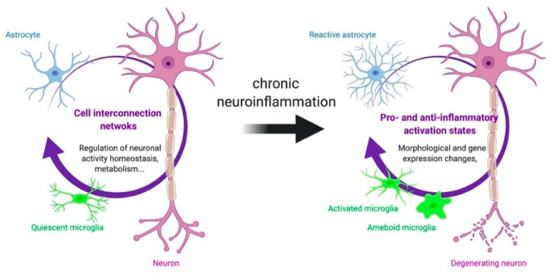
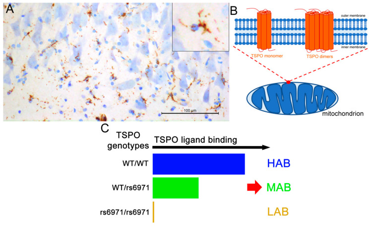
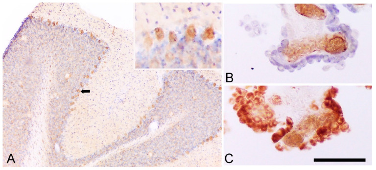
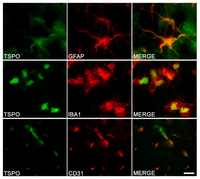
In the last decade, positron emission tomography (PET) and single-photon emission computed tomography (SPECT) in in vivo imaging has attempted to demonstrate the presence of neuroinflammatory reactions by measuring the 18 kDa translocator protein (TSPO) expression in many diseases of the central nervous system. We focus on two pathological conditions for which neuropathological studies have shown the presence of neuroinflammation, which translates in opposite in vivo expression of TSPO. Alzheimer's disease has been the most widely assessed with more than forty preclinical and clinical studies, showing overall that TSPO is upregulated in this condition, despite differences in the topography of this increase, its time-course and the associated cell types. In the case of schizophrenia, a reduction of TSPO has instead been observed, though the evidence remains scarce and contradictory. This review focuses on the key characteristics of TSPO as a biomarker of neuroinflammation in vivo, namely, on the cellular origin of the variations in its expression, on its possible biological/pathological role and on its variations across disease phases.

Keywords: Alzheimer’s disease; TSPO; astrocytes; microglia; schizophrenia.


Conflict of interest statement

The author(s) declared no conflicts of interest

Figures

Figure 1
Figure 1
Interactions between neurons, astrocytes and microglia in health and disease. In a healthy brain, the connectivity between neurons, astrocytes and microglia maintains homeostasis. In brain disease, specific morphological and functional alterations of glial cells could lead to brain degeneration.
Figure 2
Figure 2
Overview of the 18 kDa translocator protein (TSPO). (A) TSPO-immunoreactivity in the human CA1 subregion of the hippocampus. Staining looks like microglial morphology. (B) Schematic representation of TSPO monomers and dimers within the mitochondria. Homomultimeric forms exist (not shown). (C) Impact of the human TSPO rs6971 polymorphism on TSPO ligand binding and resulting population classification in HAB (high-affinity binder), MAB (mix affinity binder) and LAB (low-affinity binder). Scale bar: 100 μm.
Figure 3
Figure 3
TSPO is expressed by Purkinje and choroid plexus cells. (A) The immunoreactivity of rat cerebellum to TSPO (with cresyl violet counterstaining) underlines a positive staining in Purkinje cells (the black arrow indicates an example and insert), as demonstrated in mouse cerebellum [102]. (B) Immunoreactivity of human choroid plexus cells to CD31 (endothelial cells) with cresyl violet counterstaining (epithelial cells). (C) Immunoreactivity of human choroid plexus cells to TSPO underline both epithelial and endothelial cells positive for TSPO (as previously published in Reference [101]). Scale bar: 250 μm.
Figure 4
Figure 4
TSPO in different cell types of the hippocampus in TgF344-AD rats. Double-immunostaining was performed to detect TSPO (left column) and specific marker of astrocytes (GFAP), microglia (IBA1) and endothelial cells (CD31). Merge images demonstrate the colocalization of TSPO with astrocytes, microglia and endothelial cells. Scale bar: 10 μm. Adapted from Reference [122].
Figure 5
Figure 5
TSPO is increased in astrocytes and microglial cells in the frontal and temporal cortex of Alzheimer’s disease subjects. (AC) The [125I]CLINDE binding is used to assess TSPO density in the frontal and the temporal cortex of control (green) and AD (blue) samples. Radioactivity was determined in microglia (CD45+ cells), astrocytes (GLT1+ cells) and endothelial cells (CD31+ cells) and expressed as % injected dose (ID) /g of tissue. (D) The number of microglial cells sorted. ★: p < 0.05. Adapted from References [122,129].
Figure 6
Figure 6
Relationship between TSPO density and amyloid load in the hippocampus of 3xTg-AD mice and TgF344-AD rats. TSPO density and amyloid load were determined by in situ autoradiography with [125I]CLINDE and [125I]DRM106 in 3xTg-AD mice (A,B) and TgF344-AD rats (C,D). A positive correlation was observed in the dorsal hippocampus (A,C) but not in the ventral hippocampus (B,D). r = Spearman’s correlation coefficient. Mouse data were published in Reference [101], and rat data are personal observations.

References

    1. Bauer M.E., Teixeira A.L. Inflammation in psychiatric disorders: What comes first? Ann. NY Acad. Sci. 2019;1437:57–67. doi: 10.1111/nyas.13712. - DOI - PubMed
    1. Guilarte T.R. TSPO in diverse CNS pathologies and psychiatric disease: A critical review and a way forward. Pharmacol. Ther. 2018;194:44–58. doi: 10.1016/j.pharmthera.2018.09.003. - DOI - PMC - PubMed
    1. Brosseron F., Traschutz A., Widmann C.N., Kummer M.P., Tacik P., Santarelli F., Jessen F., Heneka M.T. Characterization and clinical use of inflammatory cerebrospinal fluid protein markers in Alzheimer’s disease. Alzheimer’s Res. Ther. 2018;10:25. doi: 10.1186/s13195-018-0353-3. - DOI - PMC - PubMed
    1. Lai K.S.P., Liu C.S., Rau A., Lanctot K.L., Kohler C.A., Pakosh M., Carvalho A.F., Herrmann N. Peripheral inflammatory markers in Alzheimer’s disease: A systematic review and meta-analysis of 175 studies. J. Neurol. Neurosurg. Psychiatry. 2017;88:876–882. doi: 10.1136/jnnp-2017-316201. - DOI - PubMed
    1. Misiak B., Frydecka D., Stanczykiewicz B., Samochowiec J. Editorial: Peripheral markers of immune response in major psychiatric disorders: Where are we now and where do we want to be? Front. Psychiatry. 2019;10:5. doi: 10.3389/fpsyt.2019.00005. - DOI - PMC - PubMed

Publication types