. 2020 Aug 24;10(1):14115.
doi: 10.1038/s41598-020-70514-z.

Programmed death ligand-1 (PD-L1) expression in meningioma; prognostic significance and its association with hypoxia and NFKB2 expression

Affiliations

Programmed death ligand-1 (PD-L1) expression in meningioma; prognostic significance and its association with hypoxia and NFKB2 expression

Shirin Karimi et al. Sci Rep. .

Abstract

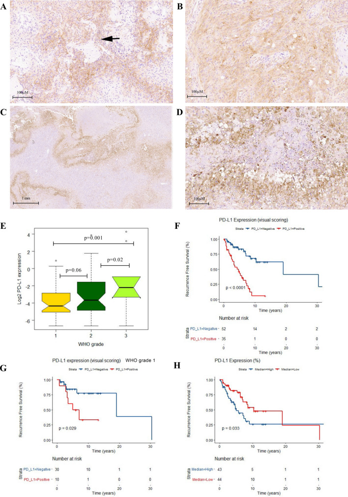
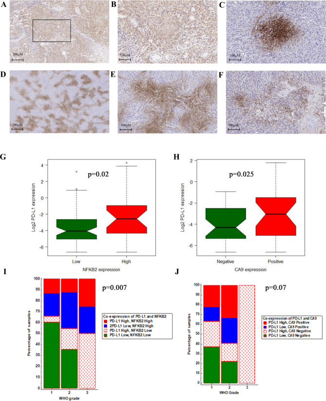
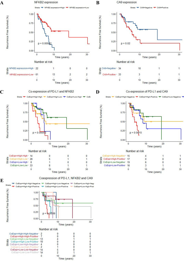
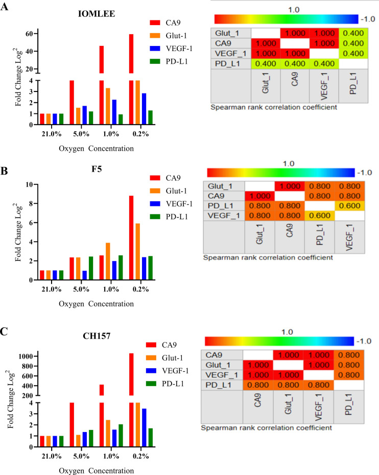
Management of clinically aggressive meningiomas is a considerable challenge. PD-L1 induced immune suppression has increasingly gained attention in clinical management of cancer; however, to date, the clinical significance and regulatory mechanisms of PD-L1 in meningioma is not yet fully characterized. We sought to characterize PD-L1 expression in meningioma and elucidate its regulatory mechanisms. Immunohistochemical staining of PD-L1 expression in meningiomas showed 43% positivity in both tumor and immune cells and we observed intra and inter tumoral heterogeneity. Univariate and multivariate analyses confirmed that PD-L1 protein expression is an independent prognostic marker for worse recurrence free survival in meningioma. Furthermore, our transcriptomic analysis revealed a strong association between PD-L1 expression and that of NFKB2 and carbonic anhydrase 9 (CA9). We also demonstrated that both of these markers, when co-expressed with PD-L1, predict tumor progression. Our studies on several meningioma cell lines cultured in hypoxic conditions validated the association of CA9 and PD-L1 expression. Here we show the clinical significance of PD-L1 in meningioma as a marker that can predict tumor recurrence. We also show an association PD-L1 expression with NFKB2 expression and its induction under hypoxic conditions. These findings may open new avenues of molecular investigation in pathogenesis of meningioma.


Conflict of interest statement

The authors declare no competing interests.

Figures

Figure 1
Figure 1
PD-L1 expression in meningioma. Representative images from heterogeneous PD-L1 IHC staining in WHO grade II with both tumor and lymphocytic positive membranous and cytoplasmic reactivity in perivascular areas (black arrow) with intratumoral extension in a WHO grade II (A,B), significant regional , per necrotic accentuation and intratumoral extension in a malignant meningioma (WHO GIII) (C,D). E Box plot graph (Two way ANOVA test demonstrates significant difference in means of PD-L1 expression (% positive cells) using HALO digital analysis in WHO grades. Kaplan Meier’s (KM) RFS analysis of PD-L1 expression (Visual scoring) in the meningioma cohort (F) and in the meningioma grade I patients (G). KM curve for digital HALO analysis of PD-L1 expression in the meningioma cohort (H) based on separation into two distinct risk groups high and low by median (0.08%). (R v3.3.1).
Figure 2
Figure 2
Representative images (× 20) from IHC staining of high cytoplasmic expression of NFKB2 (A,B) in a meningioma grade II. The CA9 IHC staining shows presence of regional, membranous and cytoplasmic in the tumor; distant from vascular channels in WHO grade II meningioma (C,D) and also in per necrotic areas of WHO grade III (E,F). The Box plot shows significant higher percentage of PD-L1 positive cells in meningioma with high NFKB2 expression (G). The same analysis in primary meningiomas indicates higher percentage of PD-L1 in positive CA9 meningioma cases (H). Chi-squared analysis demonstrated the positive association of WHO grade and co-expression of NFKB2 and PD-L1 in the meningioma cohort (I). The same analysis for WHO grade and co-expression of CA9 and PD-L1 (J). (R v3.3.1, MedCalc).
Figure 3
Figure 3
KM curves confirmed the prognostic significance for high NFKB2 expression and CA9 positivity in meningioma cases to predict worse RFS (A,B). KM analysis of predictive significance expression of these two proteins with PD-L1 in the meningioma cohort (C,D). E KM survival analysis of the IHC expressions of PD-L1, NFKB2 and CA9 in the meningioma cohort: High expression of PD-L1, NFKB2 combined with positive immunoreactivity for CA9 is associated with increased risk for tumor recurrence. (R v3.3.1).
Figure 4
Figure 4
A–C The Bar graphs and spearman’s correlation demonstrate significant positive correlation of the mRNA expression (log2 transformation) of hypoxic markers (CA9, Glut-1 and VEGF-1) and PD-L1 in three meningioma cell lines; IOMLEE, F5 and CH157 under hypoxic conditions. (MedCalc).

References

    1. Ostrom QT, et al. CBTRUS statistical report: primary brain and other central nervous system tumors diagnosed in the United States in 2010–2014. Neuro Oncol. 2017;19:v1–v88. doi: 10.1093/neuonc/nox158. - DOI - PMC - PubMed
    1. Rogers L, et al. Meningiomas: knowledge base, treatment outcomes, and uncertainties. A RANO review. J. Neurosurg. 2015;122:4–23. doi: 10.3171/2014.7.JNS131644. - DOI - PMC - PubMed
    1. Willis J, et al. The accuracy of meningioma grading: a 10-year retrospective audit. Neuropathol. Appl. Neurobiol. 2005;31:141–149. doi: 10.1111/j.1365-2990.2004.00621.x. - DOI - PubMed
    1. Clark VE, et al. Genomic analysis of non-NF2 meningiomas reveals mutations in TRAF7, KLF4, AKT1, and SMO. Science. 2013;339:1077–1080. doi: 10.1126/science.1233009. - DOI - PMC - PubMed
    1. Clark VE, et al. Recurrent somatic mutations in POLR2A define a distinct subset of meningiomas. Nat. Genet. 2016;48:1253–1259. doi: 10.1038/ng.3651. - DOI - PMC - PubMed