. 2020 Aug 24;10(1):14106.
doi: 10.1038/s41598-020-70751-2.

Palaeontological evidence reveals convergent evolution of intervertebral joint types in amniotes

Affiliations

Palaeontological evidence reveals convergent evolution of intervertebral joint types in amniotes

Tanja Wintrich et al. Sci Rep. .

Abstract

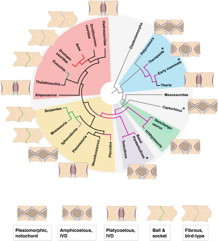
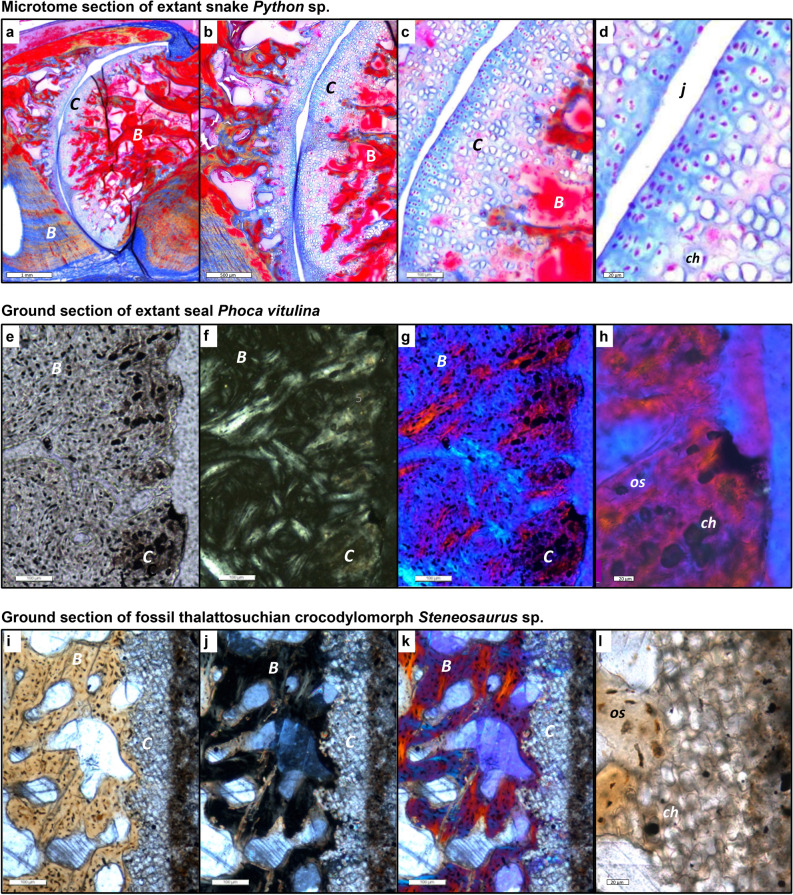
The intervertebral disc (IVD) has long been considered unique to mammals. Palaeohistological sampling of 17 mostly extinct clades across the amniote tree revealed preservation of different intervertebral soft tissue types (cartilage, probable notochord) seen in extant reptiles. The distribution of the fossilised tissues allowed us to infer the soft part anatomy of the joint. Surprisingly, we also found evidence for an IVD in fossil reptiles, including non-avian dinosaurs, ichthyosaurs, plesiosaurs, and marine crocodiles. Based on the fossil dataset, we traced the evolution of the amniote intervertebral joint through ancestral character state reconstruction. The IVD evolved at least twice, in mammals and in extinct diapsid reptiles. From this reptilian IVD, extant reptile groups and some non-avian dinosaurs independently evolved a synovial ball-and-socket joint. The unique birds dorsal intervertebral joint evolved from this dinosaur joint. The tuatara and some geckos reverted to the ancestral persisting notochord.


Conflict of interest statement

The authors declare no competing interests.

Figures

Figure 1
Figure 1
Histology of the plesiomorphic amniote joint with persisting notochord of Sphenodon punctatus NHMW 8108. The intervertebral tissues and the two articulating mid-dorsal centra illustrate the relationship between mineralised (fossilisable) tissues and soft tissues. (a) Oblique sagittal microtome section stained with hematoxylin. Note that the notochord appears discontinuous in the image because of the suboptimal plane of section. (b) Enlargement of annulus fibrosus insertion into endochondral bone of articular surface via serial cartilage. (c) Enlargement of interspinal ligament of the joint capsule inserting into periosteal bone of the centrum peripheral surface. (d) Enlargement of notochordal tissue in intervertebral space. (e) Enlargement of contact between notochordal tissue and endochondral bone of articular surface of centrum with intervening irregular cartilage. Scale bar in (a) represents 100 µm, and scale bars in (be) represent 40 µm. AF annulus fibrosus, EB endochondral bone, IC irregular cartilage, IL intervertebral ligament, N notochordal tissue, PB periosteal bone, SC serial cartilage.
Figure 2
Figure 2
Histology of mammalian and squamate intervertebral spaces. (a) Extant Phoca vitulina IGPB M 60, sagittal ground section of dorsal vertebral centrum showing part of the bony endplate in cross-polarised light with lambda filter. Note that the colors in this and the following two images do not result from histological staining but from polarised light. (b) Enlargement of area b in (a) showing a thin layer of cartilage. Note the irregular arrangement of the chondrocyte lacunae. This tissue is overlain by the nucleus pulposus (NP) of the IVD in life. (c) Enlargement of area c in (a) showing cartilage chondrocytes in inclined files (white lines) embedded in fibrous bony tissue. This represents the insertion of the annulus fibrosus (AF) of the IVD. (d) Extant Python sp. IGPB R 662, transverse microtome section of the synovial joint connecting the dorsal vertebral centrum. This joint is formed by hyaline cartilage and a thin intervertebral space filled with synovial fluid in life. (e) Fossil Mosasaurus missouriensis IGPB Goldfuß 1230, close up of the joint surface in sagittal section of a dorsal vertebral centrum. Note the globular structures arranged in files representing fossilised hyaline cartilage. (f) Enlargement of (e), white lines highlight some files. Scale bars in (a, d) represent 500 µm, scale bars in (b, c, e) represent 100 µm, and scale bar in (f) represents 20 µm. B bone tissue, C cartilage.
Figure 3
Figure 3
Histology of mesosaur, ichthyosaur and dinosaur intervertebral spaces, all fossil ground sections. (a) Stereosternum tumidum IGPB R 622, sagittal section of two articulated dorsal vertebral centra with intervertebral space, showing the notochordal amphicoelous shape and the persisting notochord. Image is in cross-polarised light with lambda filter. (b) Hadrosauridae indet. UALVP 59650. Close up of the articular surface showing obliquely arranged mineralised fibres in between poorly defined files of chondrocyte lacunae (arrows). Image is in cross-polarised light with lambda filter. (c) Stenopterygius sp. IGPB R 661 sagittal section of two articulated centra showing the amphicoelous shape. Note the differentiation of the content of the intervertebral space into a coarse into a fine fraction, probably representing the nucleus pulposus (NP) and the annulus fibrosus (AF). (d) Enlargement of the concave part of the articular surface, showing a thin layer of irregularly arranged chondrocyte lacunae, underlying the nucleus pulposus (NP). (e) Enlargement of the convex part of the articular surface, showing obliquely arranged files of chondrocyte lacunae, representing the insertion of the annulus fibrosus (AF) (arrows). Scale bar in (a) represents 500 µm, scale bar in (b) represents 100 µm, scale bar in (c) represents 2 mm, scale bars in (d, e) represent 100 µm. AF annulus fibrosus, B bone tissue, C cartilage, N notochord, NC neural canal, NP nucleus pulposus.
Figure 4
Figure 4
Ancestral state reconstruction using parsimony of the different types of dorsal intervertebral joints in the phylogeny of the higher clades of amniotes (see “Methods” and SM for details). The schematic drawings of the intervertebral joints are explained in the text. Grey branches indicate persisting notochord. Purple branches indicate an intervertebral disc between amphicoelous centra. Black branches indicate an intervertebral disc between platycoelous centra. Green branches indicate a synovial joint with hyaline cartilage in between procoelous or opisthocoelous centra. Red branches indicate a fibrous cartilage joint. Key to tissue colors: beige, bone; purple, cartilaginous endplate; light blue, AF; brown, NP; green, articular cartilage of synovial joint; grey, fibrous joint cartilage. Key to clade colors: light blue, Synapsida; light green, Ichthyosauria; light purple, Testudines; light yellow, Lepidosauromorpha; light red, Archosauromorpha. Asterisks indicate fossil taxa for which intervertebral soft tissue anatomy was inferred based on morphological and histological descriptions in the literature.
Figure 5
Figure 5
Intervertebral articular cartilage in extant and fossil amniotes and different types of sectioning and light microscopy. (ad) Microtome section stained with Azan of extant joint with cartilage of extant snake Python sp. IGPB R 662, synovial ball-and-socket joint at increasing magnifications in normal transmitted light. Blue is cartilage and other connective tissues, red is bone. Note the serial cartilage, i.e., the arrangement of the cartilage cells in files, especially in (c). (d) Cartilage layer with differentiating (left) and hypertrophied (right) cartilage cells, note the size increase from left to right and the globular shape of the hypertrophied chondrocytes, as in (h) and (l). (eh) Ground section of extant seal Phoca vitulina IGPB M 60, joint with IVD, peripheral area of articular surface. (e) Section in normal transmitted light. (f) Section in cross-polarised light. (g) Cross-polarised light and lambda filter. (h) Close-up of g. (il) Ground section of fossil thalattosuchian crocodilomorph Steneosaurus IGPB R 663, joint with IVD, peripheral area of articular surface. (i) Section in normal light. (j) Section in cross-polarised light. (k) Cross polarised light and lambda filter. (l) Close-up of (i). Note that the fossil cartilage does not show the same birefringence as the bone. B bone tissue, C cartilage, ch chondrocyte, j joint space, os osteocyte.

Similar articles

Cited by

References

    1. Romer AS. Osteology of the Reptiles. Chicago: The University of Chicago Press; 1956. p. 772.
    1. Hall BK. Bones and Cartilage. Developmental and Evolutionary Skeletal Biology. 2. San Diego: Academic Press; 2015. p. 902.
    1. Bruggeman BJ, et al. Avian intervertebral disc arises from rostral sclerotome and lacks a nucleus pulposus: implications for evolution of the vertebrate disc. Dev. Dyn. 2012;241:675–683. - PMC - PubMed
    1. Witzmann F, Schwarz-Wings D, Hampe O, Fritsch G, Asbach P. Evidence of spondyloarthropathy in the spine of a phytosaur (Reptilia: Archosauriformes) from the Late Triassic of Halberstadt Germany. PLoS ONE. 2014;9:e85511. - PMC - PubMed
    1. Schweitzer MH. Soft tissue preservation in terrestrial Mesozoic vertebrates. Annu. Rev. Earth Planet. Sci. 2011;39:187–216.

Publication types

MeSH terms