. 2020 Sep 18;15(9):2529-2538.
doi: 10.1021/acschembio.0c00564. Epub 2020 Sep 8.

Functional and Structural Insights into a Novel Promiscuous Ketoreductase of the Lugdunomycin Biosynthetic Pathway

Affiliations

Functional and Structural Insights into a Novel Promiscuous Ketoreductase of the Lugdunomycin Biosynthetic Pathway

Xiansha Xiao et al. ACS Chem Biol. .

Abstract

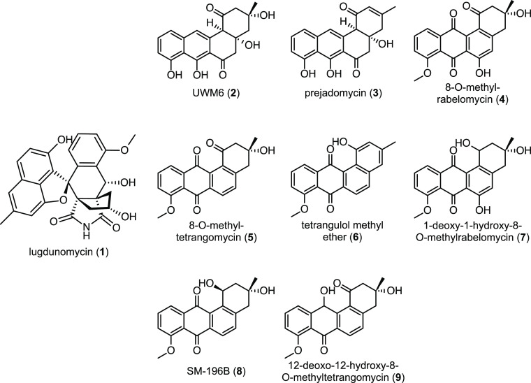
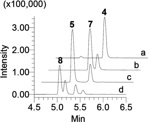
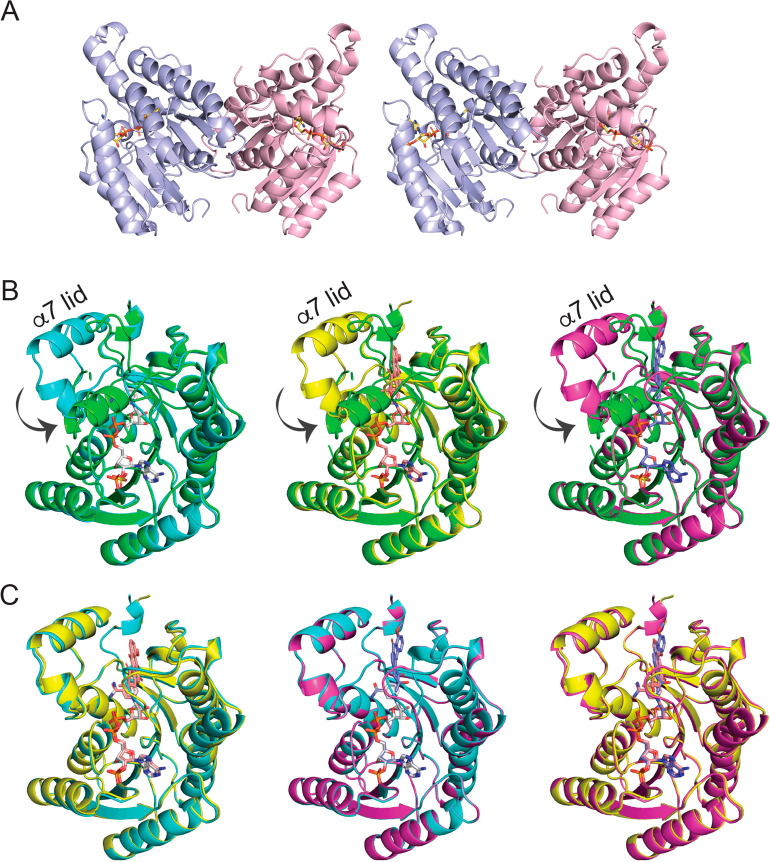
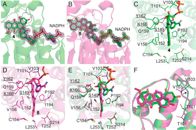
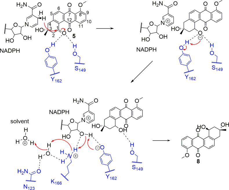
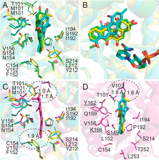
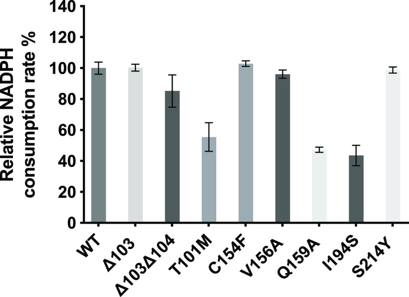
Angucyclines are a structurally diverse class of actinobacterial natural products defined by their varied polycyclic ring systems, which display a wide range of biological activities. We recently discovered lugdunomycin (1), a highly rearranged polyketide antibiotic derived from the angucycline backbone that is synthesized via several yet unexplained enzymatic reactions. Here, we show via in vivo, in vitro, and structural analysis that the promiscuous reductase LugOII catalyzes both a C6 and an unprecedented C1 ketoreduction. This then sets the stage for the subsequent C-ring cleavage that is key to the rearranged scaffolds of 1. The 1.1 Å structures of LugOII in complex with either ligand 8-O-Methylrabelomycin (4) or 8-O-Methyltetrangomycin (5) and of apoenzyme were resolved, which revealed a canonical Rossman fold and a remarkable conformational change during substrate capture and release. Mutational analysis uncovered key residues for substrate access, position, and catalysis as well as specific determinants that control its dual functionality. The insights obtained in this work hold promise for the discovery and engineering of other promiscuous reductases that may be harnessed for the generation of novel biocatalysts for chemoenzymatic applications.


Conflict of interest statement

The authors declare no competing financial interest.

Figures

Figure 1
Figure 1
Structures of the metabolites discussed in this study. Lugdunomycin (1), UWM6 (2), prejadomycin (3), 8-O-methylrabelomycin (4), 8-O-methyltetrangomycin (5), tetrangulol methyl ether (6), 1-deoxo-1-hydroxy-8-O-methylrabelomycin (7), SM-196B (8), and 12-deoxo-12-hydroxy-8-O-methyltetrangomycin (9).
Figure 2
Figure 2
Extracted-ion chromatogram (XIC) overlay of the ion peaks of all the related compounds from the in vitro reactions. Enzymatic reactions: (a) 4; (b) 4 + LugOII; (c) 5; (d) 5 + LugOII. The experiments were independently repeated three times with similar results.
Figure 3
Figure 3
Dimeric arrangement of LugOII structures and observed conformational changes. (A) Stereoscopic view of NADPH-bound LugOII in dimeric form. (B) Unliganded LugOII (green), LugOII/NADPH (cyan), LugOII/NADPH/4 (yellow), and LugOII/NADPH/5 (magenta) structures are superimposed. α6, α7, and the loop region between them serve as a lid, which turn around 180° and then rotate 90° toward the binding site of 5. (C) Alignment of LugOII/NADPH (cyan), LugOII/NADPH/4 (yellow), and LugOII/NADPH/5 (magenta) structures. NADPH, 4, and 5 are displayed in sticks.
Figure 4
Figure 4
Active site of LugOII. (A, B) 2FoFc omit maps contoured at the 1σ level corresponding to ligands 4 and 5 and cofactor NADPH. The missing density for 4 is highlighted with a black circle. (C, D) Key residues that surround the binding site of 4 and 5. Catalytic residues Ser149, Tyr162, and Lys166 are underlined, and their distances (within 3.2 Å) to the ligand and cofactor are dashed. (E, F) Superposition of the two substrates 4 and 5 bounded to LugOII. (E) Top view of the active pocket. (F) Side view of the two aligned substrate structures. Major differences are found in the orientations of the two substrates and the movement of the α4−β4 loop that are close to the A-rings of the two substrates. See also Figure S6.
Figure 5
Figure 5
Postulated catalytic mechanism for the LugOII-catalyzed C1 reduction and proton relay. The reaction is initiated by proton transfer from the hydroxyl group of Tyr162 to the carbonyl group of 5, followed by a hydride transfer to the C1 position of 5. The catalytic triad (Lys166, Tyr162, and Ser149) and N123 are highlighted in blue.
Figure 6
Figure 6
Superposition of LugOII (green for 4 and magenta for 5), LanV (cyan, PDB entry, 4KWI), and UrdMred (yellow, PDB entry, 4OSP) reveals major differences in the active sites. (A, B) LugOII/4, LanV, and UrdMred structures are aligned. (C) 5 is superposed in the active pocket of LanV and UrdMred. Clashes were seen in two regions (highlighted by circles). One region is the extra loop between α4 and β4 in which Thr101 of LugOII is replaced by Met101 of LanV and UrdMred. The other region represents residues Cys154 and Ser214 that are near the D-ring binding site. They were substituted by Val152 and Tyr212 in LanV and Phe152 and Leu212 in UrdMred, respectively. (D) 11-Deoxylandomycinone of LanV and rabelomycin of UrdMred are superposed in the LugOII active pocket, where Val103 of LugOII clashes with the 3-methyl and 3-hydroxyl groups of the A-ring, respectively.
Figure 7
Figure 7
Enzymatic activity of LugOII variants. The columns represent the relative activity of LugOII variants compared to that of the wild-type enzyme, based on the consumption rate of NADPH by measuring the UV absorbance at 340 nm. Reactions were carried out using compound 5 as the substrate. The experiments were independently repeated three times with similar results.

Similar articles

Cited by

References

    1. Kharel M. K.; Pahari P.; Shepherd M. D.; Tibrewal N.; Nybo S. E.; Shaaban K. A.; Rohr J. (2012) Angucyclines: biosynthesis, mode-of-action, new natural products, and synthesis. Nat. Prod. Rep. 29, 264–325. 10.1039/C1NP00068C. - DOI - PMC - PubMed
    1. Ostash B.; Korynevska A.; Stoika R.; Fedorenko V. (2009) Chemistry and biology of landomycins, an expanding family of polyketide natural products. Mini-Rev. Med. Chem. 9, 1040–1051. 10.2174/138955709788922593. - DOI - PubMed
    1. Drautz H.; Zahner H.; Rohr J.; Zeeck A. (1986) Metabolic products of microorganisms. 234. Urdamycins, new angucycline antibiotics from Streptomyces fradiae. I. Isolation, characterization and biological properties. J. Antibiot. 39, 1657–1669. 10.7164/antibiotics.39.1657. - DOI - PubMed
    1. Yang K.; Han L.; Vining L. C. (1995) Regulation of jadomycin B production in Streptomyces venezuelae ISP5230: involvement of a repressor gene, jadR2. J. Bacteriol. 177, 6111–6117. 10.1128/JB.177.21.6111-6117.1995. - DOI - PMC - PubMed
    1. Takahashi K.; Yoshida M.; Tomita F.; Shirahata K. (1981) Gilvocarcins, new antitumor antibiotics. 2. Structural elucidation. J. Antibiot. 34, 271–275. 10.7164/antibiotics.34.271. - DOI - PubMed

Publication types

MeSH terms