

Discrepancies in digital hematopathology diagnoses for consultation and expert panel analysis
- PMID: 32840673
- PMCID: PMC7973407
- DOI: 10.1007/s00428-020-02907-4
Discrepancies in digital hematopathology diagnoses for consultation and expert panel analysis
Abstract
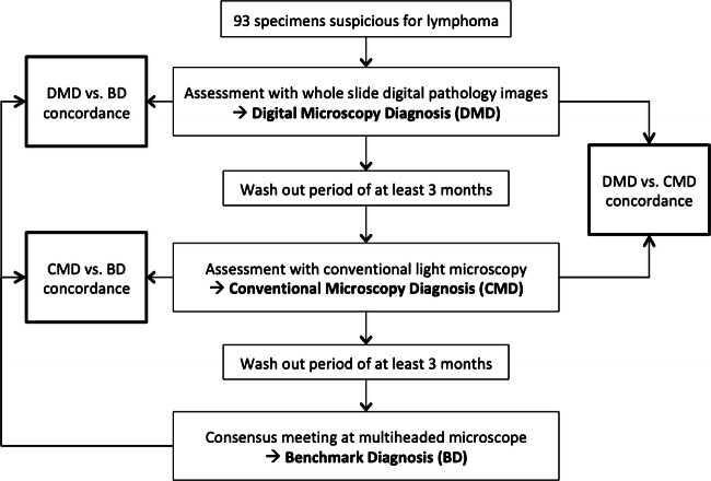
Digital pathology with whole-slide imaging (WSI) has a large potential to make the process of expert consultation and expert panel diagnosis more rapid and more efficient. However, comparison with the current methods is necessary for validation of the technique. In this study, we determined if digital assessment of whole-slide images of hematopathology specimens with a focus on the assessment of lymphoma can be used for consultation and panel diagnostics. Ninety-three histological specimens with a suspicion for lymphoma were assessed both with conventional microscopy and digital microscopy with a wash out period between assessments. A consensus diagnosis was based on full concordance between the pathologists or, in case of discordances, was reached at a joint session at a multi-headed microscope. In 81% of the cases, there was a full concordance between digital and light microscopical assessment for all three pathologists. Discordances between conventional microscopy and digital pathology were present in 3% of assessments. In comparison with the consensus diagnosis, discordant diagnoses were made in 5 cases with digital microscopy and in 3 cases with light microscopy. The reported level of confidence and need for additional investigations were similar between assessment by conventional and by digital microscopy. In conclusion, the performance of assessment by digital pathology is in general comparable with that of conventional light microscopy and pathologists feel confident using digital pathology for this subspecialty.
Keywords: Consultation; Digital pathology; Hematopathology; Lymphoma.
Conflict of interest statement
The authors declare that they have no conflicts of interest.
Figures
References
-
- LaCasce AS, Kho ME, Friedberg JW, Niland JC, Abel GA, Rodriguez MA, Czuczman MS, Millenson MM, Zelenetz AD, Weeks JC. Comparison of referring and final pathology for patients with non-Hodgkin’s lymphoma in the National Comprehensive Cancer Network. J Clin Oncol. 2008;26(31):5107–5112. doi: 10.1200/JCO.2008.16.4061. - DOI - PMC - PubMed
-
- Strobbe L, van der Schans SA, Heijker S, Meijer JW, Mattijssen EJ, Mandigers CM, de Kievit IM, Raemaekers JM, Hebeda KM, van Krieken JH. Evaluation of a panel of expert pathologists: review of the diagnosis and histological classification of Hodgkin and non-Hodgkin lymphomas in a population-based cancer registry. Leuk Lymphoma. 2014;55(5):1018–1022. doi: 10.3109/10428194.2013.827787. - DOI - PubMed
-
- van Diest PJ, Huisman A, van Ekris J, Meijer J, Willems S, Hofhuis H, Verbeek X, van der Wel M, Vos S, Leguit R, van den Brand M, Hebeda K, Grunberg K. Pathology image exchange: the Dutch digital pathology platform for exchange of whole-slide images for efficient teleconsultation, telerevision, and virtual expert panels. JCO Clin Cancer Inform. 2019;3:1–7. doi: 10.1200/CCI.18.00146. - DOI - PMC - PubMed
-
- Snead DR, Tsang YW, Meskiri A, Kimani PK, Crossman R, Rajpoot NM, Blessing E, Chen K, Gopalakrishnan K, Matthews P, Momtahan N, Read-Jones S, Sah S, Simmons E, Sinha B, Suortamo S, Yeo Y, El Daly H, Cree IA. Validation of digital pathology imaging for primary histopathological diagnosis. Histopathology. 2016;68(7):1063–1072. doi: 10.1111/his.12879. - DOI - PubMed
Publication types
MeSH terms
LinkOut - more resources
Full Text Sources
Medical
