

Do different cone beam computed tomography exposure protocols influence subjective image quality prior to and after root canal treatment?
- PMID: 32840680
- PMCID: PMC7966640
- DOI: 10.1007/s00784-020-03524-w
Do different cone beam computed tomography exposure protocols influence subjective image quality prior to and after root canal treatment?
Abstract
Objectives: The current study aimed to evaluate different CBCT exposure protocols and influencing factors affecting the subjective image quality of scans taken for endodontic indications.
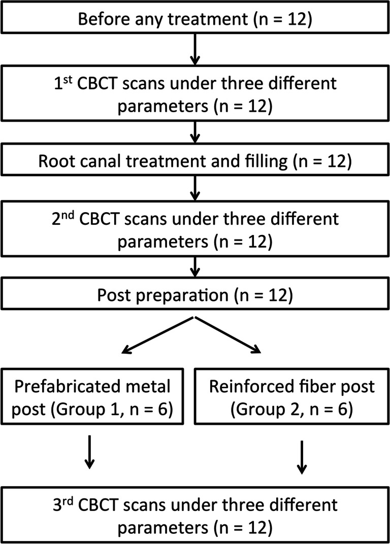
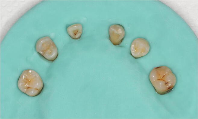
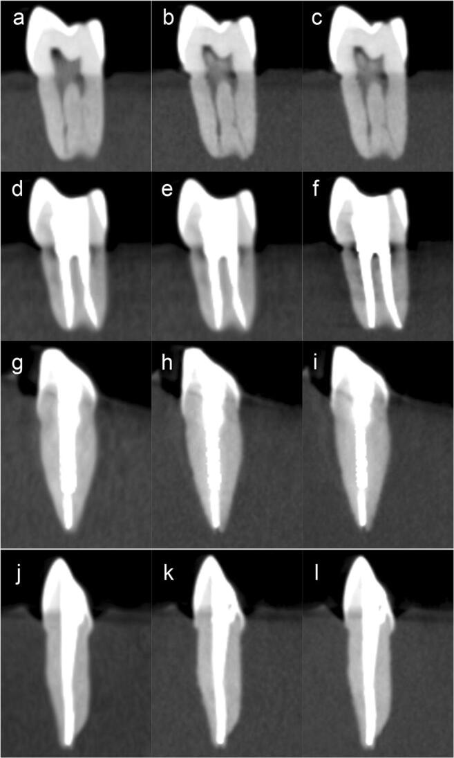
Materials and methods: Twelve extracted teeth, comprising of two sets of maxillary molars, premolars, canines and incisors, mandibular premolars, and molars, were endodontically treated, and either received a fiber or metal post. The teeth were scanned by CBCT imaging before and after root canal treatment, and after post insertion. Each scan was performed thrice, using an ultra low dose (ULD), standard (SM), and high-resolution mode (HR), respectively. Twelve observers-4 endodontists, 4 periodontists, and 4 radiologists-assessed the subjective image quality using visual analogue scales (VAS). Potential influencing factors were evaluated including acquisition mode, observer specialty, stage of treatment, type of post, and type of tooth, using one-way ANOVA and T test.
Results: Teeth scanned with the ULD had the highest average VAS score (72.5), followed by HR (70.2), and SM (69.0) for values pooled from all teeth and observers. CBCT acquisition mode was not a significant influencing factor on the VAS scores. Observer specialty, stage of treatment, type of post, and type of tooth were significant influencing factors.
Conclusions: Based on the present in vitro data, a low-dose CBCT mode seems not to negatively affect the perception of image quality.
Clinical relevance: The findings from this in vitro study demonstrate that a low-dose CBCT mode might have potential for diagnostics prior to or following endodontic treatment.
Keywords: Cone beam computed tomography; Dose optimization; Image quality; Low dose; Root canal treatment.
Conflict of interest statement
The authors declare that they have no conflict of interest.
Figures
References
MeSH terms
Grants and funding
LinkOut - more resources
Full Text Sources
