. 2020 Oct 2;5(19):e142032.
doi: 10.1172/jci.insight.142032.

Human angiotensin-converting enzyme 2 transgenic mice infected with SARS-CoV-2 develop severe and fatal respiratory disease

Affiliations

Human angiotensin-converting enzyme 2 transgenic mice infected with SARS-CoV-2 develop severe and fatal respiratory disease

Joseph W Golden et al. JCI Insight. .

Abstract

The emergence of SARS-CoV-2 has created an international health crisis, and small animal models mirroring SARS-CoV-2 human disease are essential for medical countermeasure (MCM) development. Mice are refractory to SARS-CoV-2 infection owing to low-affinity binding to the murine angiotensin-converting enzyme 2 (ACE2) protein. Here, we evaluated the pathogenesis of SARS-CoV-2 in male and female mice expressing the human ACE2 gene under the control of the keratin 18 promoter (K18). In contrast to nontransgenic mice, intranasal exposure of K18-hACE2 animals to 2 different doses of SARS-CoV-2 resulted in acute disease, including weight loss, lung injury, brain infection, and lethality. Vasculitis was the most prominent finding in the lungs of infected mice. Transcriptomic analysis from lungs of infected animals showed increases in transcripts involved in lung injury and inflammatory cytokines. In the low-dose challenge groups, there was a survival advantage in the female mice, with 60% surviving infection, whereas all male mice succumbed to disease. Male mice that succumbed to disease had higher levels of inflammatory transcripts compared with female mice. To our knowledge, this is the first highly lethal murine infection model for SARS-CoV-2 and should be valuable for the study of SARS-CoV-2 pathogenesis and for the assessment of MCMs.

Keywords: COVID-19; Infectious disease; Molecular pathology; Mouse models.


Conflict of interest statement

Conflict of interest: The authors have declared that no conflict of interest exists.

Figures

Figure 1
Figure 1. SARS-CoV-2 infection in K18-hACE2 transgenic mice.
(A) Male and female K18-hACE2 transgenic mice (day 0–3, n = 7/group; day 3+, n = 5/group) were infected with 2 × 10⁴ PFU or 2 × 10³ PFU of SARS-CoV-2 by the IN route. C57BL/6 and RAG2 KO mice (day 0–3, n = 8/ group; day 3+, n = 5/group) were infected with 2 × 10⁴ PFU by the IN route. Survival and weight loss (± SEM) were monitored and plotted using Prism software. (B) Titers in lung (n = 2 mice/group) were examined on day 3 by qRT-PCR. Mean titers ± SEM of the genome molecules of viral RNA/mL were graphed. (C) Titers in lungs of individual K18-hACE2 mice. Numbers above bars denote day of death. Colors represent the 4 groups. (D) Monocyte chemoattractants and inflammatory cytokines were measured from the serum of SARS-CoV-3– infected mice on day 3 or at the time of euthanasia using a multiplex system. Mice from each group are aggregated from samples taken on day 3 (blue symbols) and when mice were euthanized (black symbols).
Figure 2
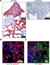
Figure 2. Infection of SARS-CoV-2 in the lung of K18-hACE2 transgenic mice.
(A) Representative ISH images showing the presence of SARS-CoV-2 RNA (red) in the lungs of infected mice at low and high magnification or uninfected mice. Cells were counterstained with hematoxylin (blue). (B) Costaining for viral spike protein (green) and E-cadherin (red) in infected lung tissues using IFA. Arrows point to double-positive cells. Nuclei are stained with DAPI (blue). (C) Costaining of viral nucleoprotein and the macrophage marker CD68 (red) in infected lungs using IFA. Arrows denote double-positive cells. Nuclei are stained with DAPI (blue).
Figure 3
Figure 3. SARS-CoV-2 infection causes respiratory damage in K18-hACE2 mice.
(AD) Representative H&E staining of lungs in infected C57BL/6 mice (A) or K18-hACE2-infected mice (BD). Numerous fibrin thrombi (black arrows) filling the lumen of small-to-intermediate size vessels (B) adjacent to a normal bronchus and surrounded by minimally inflamed, congested, and collapsed alveolar septa. (C) Extensive area of lung consolidation with inflammation and expansion of alveolar septa, exudation of fibrin and edema into alveolar lumina, and infiltration of vessel walls and perivascular area by numerous mononuclear inflammatory cells (arrows), disrupting/obscuring vessel architecture (vasculitis). (D) Extensive area of consolidated lung showing type II pneumocyte hyperplasia (arrowhead) and rare multinucleate cells (black arrow). (E) H&E and ISH staining of infected mouse lung showing vasculitis with absence of viral RNA in the affected vessel walls; note there is viral RNA (red) in the adjacent alveolar septa. Highlighted vessels (broken black circles). (F) TUNEL staining of infected and uninfected K18-hACE2 mouse lungs. TUNEL (green) was performed as indicted in Methods. Cell nuclei stained with DAPI (blue). (G) Ki-67 staining (red) in infected and uninfected K18-hACE2 mouse lungs. Nuclei stained with DAPI (blue).
Figure 4
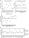
Figure 4. Transcriptional activation in SARS-CoV-2–infected lungs.
Transcriptional activation in lung homogenates from infected K18-hACE2 (male and female) and C57BL/6 (female) mice were examined by NanoString. (A) Log2 fold changes in gene expression levels of selected genes categorized by group vs. infected C57BL/6 mice were graphed with SD. All graphed transcripts had a P value of less than 0.05. (B) Differential gene expression (log2 changes) between infected male and female K18-hACE2 mice. All graphed transcripts had a P value of less than 0.05.
Figure 5
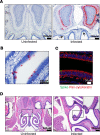
Figure 5. Infection of the nasal cavity and olfactory bulb in K18-hACE2 mice.
(A) ISH labeling for SARS-CoV-2 RNA in a coronal section of the head, including the caudal aspect of the nasal cavity and olfactory bulb. Within the olfactory bulb, a strong positive signal is present in the glomerular layer, external plexiform layer, mitral cell layer, and internal plexiform layer, with multifocal positivity in the granular cell layer in the olfactory bulb hemisphere at right. Low numbers of cells within the olfactory epithelium lining the dorsal nasal meatus have a positive ISH signal (arrows). Cells were counterstained with hematoxylin (blue). (B) ISH labeling for SARS-CoV-2 RNA in the olfactory epithelium. Cells were counterstained with hematoxylin (blue). (C) IFA of olfactory epithelium stained for SARS-CoV-2 spike (green) and Pan-cytokeratin (red). Nuclei were stained with DAPI (blue). (D) Representative H&E staining of the nasal cavity, including olfactory epithelium and olfactory bulb from uninfected or infected mice. In the infected mouse, there is a focal area of olfactory epithelium atrophy on a nasal turbinate located in the lateral nasal meatus. Inset, indicated area of detail under increased magnification.
Figure 6
Figure 6. Neuropathogenesis of SARS-CoV-2 in K18-hACE2 transgenic and nontransgenic mice.
ISH detection of for SARS-CoV-2 RNA in uninfected (A) and infected mice (B and C) in a coronal section of brain demonstrating a strong positive signal within neurons of thalamic nuclei. The boxed area is shown at increased magnification (right) (C). Note the absence of a positive signal from the vessel at center right (C) where the vessel wall and perivascular space are infiltrated by mononuclear inflammatory cells. (DG) Representative H&E staining of uninfected (D) or infected (EG) mice. Perivascular hemorrhage extending into the adjacent neuropil (E), in the region of the rostral cerebral cortex. Two small caliber vessels (F) in the thalamus with fibrin thrombi (arrows), with mild microgliosis and some perivascular hemorrhage (arrowhead). The walls of small-to-intermediate size vessels and perivascular spaces (G) are multifocally expanded/obscured by mononuclear inflammatory cells and increased numbers of glial cells. (H) Detection of Iba-1 and GFAP, markers for microgliosis and astrogliosis, respectively, in uninfected or infected brain sections using IFA. Nuclei were stained with DAPI (blue). (I) Costaining for astrocyte marker GFAP (red) or neuron marker NeuN (red) with SARS-CoV-2 spike protein (green). Spike protein was predominantly detected in NeuN+ neurons. Nuclei were stained with DAPI (blue).

References

    1. Chen N, et al. Epidemiological and clinical characteristics of 99 cases of 2019 novel coronavirus pneumonia in Wuhan, China: a descriptive study. Lancet. 2020;395(10223):507–513. - PMC - PubMed
    1. Zhou P, et al. A pneumonia outbreak associated with a new coronavirus of probable bat origin. Nature. 2020;579(7798):270–273. doi: 10.1038/s41586-020-2012-7. - DOI - PMC - PubMed
    1. Yang X, et al. Clinical course and outcomes of critically ill patients with SARS-CoV-2 pneumonia in Wuhan, China: a single-centered, retrospective, observational study. Lancet Respir Med. 2020;8(5):475–481. doi: 10.1016/S2213-2600(20)30079-5. - DOI - PMC - PubMed
    1. Barnes BJ, et al. Targeting potential drivers of COVID-19: neutrophil extracellular traps. J Exp Med. 2020;217(6):e20200652. doi: 10.1084/jem.20200652. - DOI - PMC - PubMed
    1. Salimi S, Hamlyn JM. COVID-19 and crosstalk with the hallmarks of aging. J Gerontol A Biol Sci Med Sci. 2020;null(9):glaa149. - PMC - PubMed

Publication types

MeSH terms