. 2020 Aug 25;18(8):e3000788.
doi: 10.1371/journal.pbio.3000788. eCollection 2020 Aug.

Glycan cross-feeding supports mutualism between Fusobacterium and the vaginal microbiota

Affiliations

Glycan cross-feeding supports mutualism between Fusobacterium and the vaginal microbiota

Kavita Agarwal et al. PLoS Biol. .

Abstract

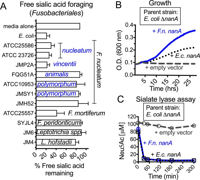
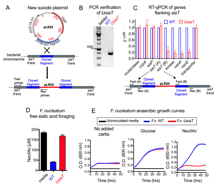
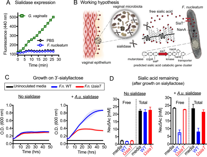
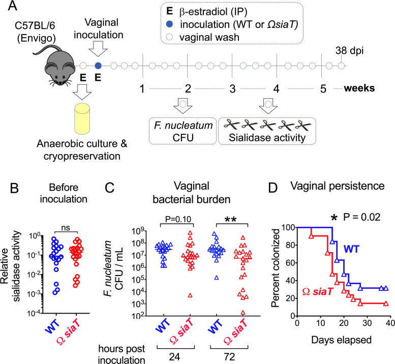
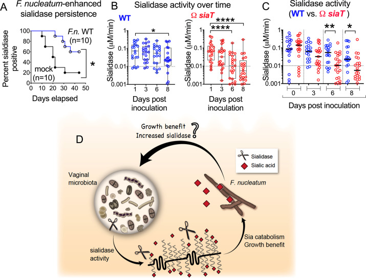
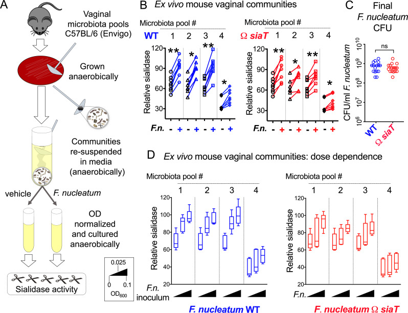
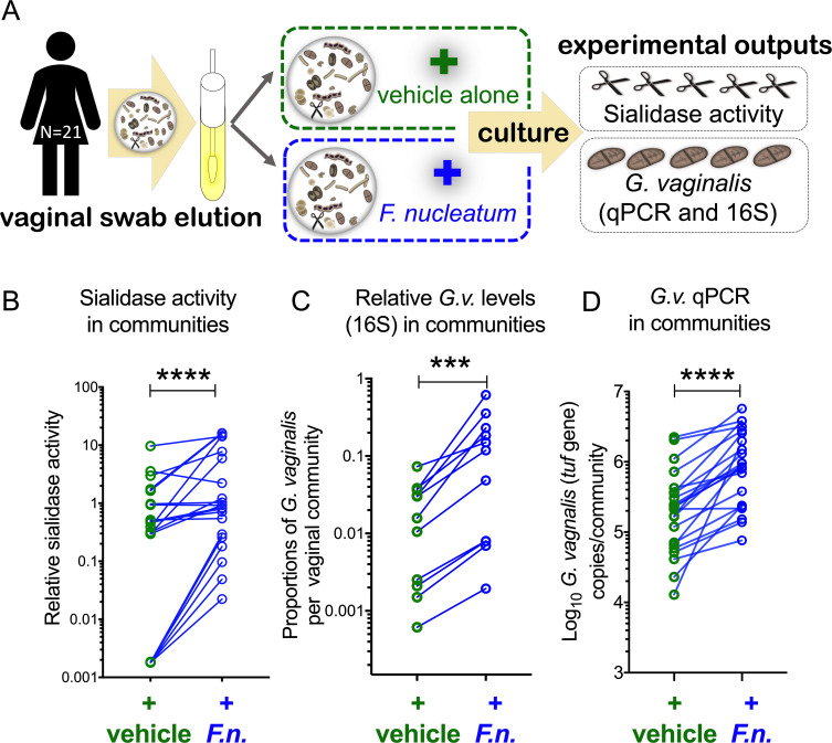
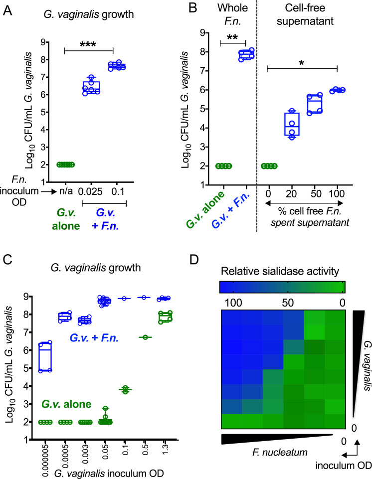
Women with bacterial vaginosis (BV), an imbalance of the vaginal microbiome, are more likely to be colonized by potential pathogens such as Fusobacterium nucleatum, a bacterium linked with intrauterine infection and preterm birth. However, the conditions and mechanisms supporting pathogen colonization during vaginal dysbiosis remain obscure. We demonstrate that sialidase activity, a diagnostic feature of BV, promoted F. nucleatum foraging and growth on mammalian sialoglycans, a nutrient resource that was otherwise inaccessible because of the lack of endogenous F. nucleatum sialidase. In mice with sialidase-producing vaginal microbiotas, mutant F. nucleatum unable to consume sialic acids was impaired in vaginal colonization. These experiments in mice also led to the discovery that F. nucleatum may also "give back" to the community by reinforcing sialidase activity, a biochemical feature of human dysbiosis. Using human vaginal bacterial communities, we show that F. nucleatum supported robust outgrowth of Gardnerella vaginalis, a major sialidase producer and one of the most abundant organisms in BV. These results illustrate that mutually beneficial relationships between vaginal bacteria support pathogen colonization and may help maintain features of dysbiosis. These findings challenge the simplistic dogma that the mere absence of "healthy" lactobacilli is the sole mechanism that creates a permissive environment for pathogens during vaginal dysbiosis. Given the ubiquity of F. nucleatum in the human mouth, these studies also suggest a possible mechanism underlying links between vaginal dysbiosis and oral sex.


Conflict of interest statement

The authors declare that they have no relevant conflicts of interest.

Figures

Fig 1
Fig 1. Analysis of sialic acid utilization by Fusobacteria and functional evidence of F. nucleatum-encoded sialate lyase (nanA).
(A) Amount of sialic acid remaining in the growth medium 24–48 h postinoculation of different Fusobacteria (as indicated) or uninoculated media control. Subspecies of F. nucleatum are indicated in blue text. All Fusobacteria were grown in supplemented Columbia medium with free sialic acid added (Neu5Ac, 100 μM)—medium that was also used as uninoculated control. E. coli (MG1655), a known sialic acid consumer, cultured in LB medium with added free sialic acid (Neu5Ac, 100 μM) was included as a positive control. Error bars shown are standard deviation of the mean from 3 independent experiments. (B–C) E. coli MG1655 ΔnanA was complemented with empty vector, E. coli nanA or putative nanA from F. nucleatum ATCC23726 (GenBank accession EFG95907). Data shown are representative of 3 independent experiments. (B) Growth was assessed in minimal media with sialic acid by measuring absorbance at 600 nm. (C) Lysates of the E. coli nanA mutant with empty vector or the complemented strain were incubated with sialic acid (Neu5Ac), and its disappearance was monitored over time by fluorescent derivatization (with DMB) followed by HPLC. The underlying numerical data for this figure can be found in S1 Data. Strain names: FQG51A, JM6, JM4, JMH52, JMP2A, JMSY1, SYJL4. Description of bacterial strains can be found in S1 Table. ATCC, American Type Culture Collection; DMB, 1,2-diamino-4,5-methylenedioxybenzene; E.c., E. coli; F.n., F. nucleatum; HPLC, high-performance liquid chromatography; LB, lysogeny broth; nanA, N-acetylneuraminate lyase; Neu5Ac, N-acetylneuraminic acid; OD, optical density.
Fig 2
Fig 2. Insertional mutagenesis to disrupt sialic acid catabolic pathway in F.n. ATCC23726 and growth analysis in nutrient-limited media.
(A) Schematic showing integration of plasmid containing siaT insert (pLR25) into the F.n. chromosome. Positions of Fwd and Rev primers used for confirming integration of plasmid are also indicated. (B) Agarose gel image with the expected PCR product confirming integration of plasmid into the siaT locus. (C) RT-qPCR analysis of genes flanking siaT in F.n. shows low expression of transcripts of putative sialic acid catabolism genes downstream of the plasmid insertion site. Difference in expression of each gene between F.n. WT versus ΩsiaT was analyzed by the ΔΔCt method using the 16S rRNA gene for normalization. Fwd and Rev primer binding sites are indicated by arrows in the schematic below the graph. (D) Functional assessment of the WT and ΩsiaT strains confirms disruption of sialic acid catabolism. Shown is the concentration of sialic acid (Neu5Ac) remaining in the medium 24 hpi. Sialic acid consumption by F.n. WT and ΩsiaT was studied in supplemented Columbia media with free sialic acid added. (E) Growth of F.n. WT and ΩsiaT in nutrient-limited media with no added carbohydrates (carbs) or with the indicated carbohydrates added. All data shown are representative of 2 or more independent experiments. The underlying numerical data for this figure can be found in S1 Data. Error bars represent standard deviation from the mean. Description of the plasmids and cloning vectors used for construction of new suicide plasmid pLR25 can be found in S1 Table. Description of the genes in the predicted sialic acid catabolic gene cluster can be found in S2 Table. ATCC, American Type Culture Collection; catP, chloramphenicol acetyltransferase; Fwd, forward; F.n., F. nucleatum; hpi, hours postinoculation; nanA, N-acetylneuraminate lyase; Neu5Ac, N-acetylneuraminic acid; Rev, reverse; RT-qPCR, reverse transcription–quantitative PCR; siaT, predicted sialic acid transporter; WT, wild type.
Fig 3
Fig 3. F.n. accesses bound sialic acid from glycan chains, but only when liberated by exogenous sialidases.
(A) Analysis of cell-associated sialidase activity of anaerobically cultured G. vaginalis JCP8151B and F.n. ATCC23726 strains using fluorogenic 4MU-Neu5Ac substrate. (B) Working hypothesis: sialidase producers in the vaginal microbial community release free sialic acids (red diamonds) from host glycoconjugates, which may be accessed by F.n., which does not produce sialidase. More information about F.n. genes shown in the predicted sialic acid catabolic gene cluster and the enzymes they encode can be found in S2 Table. (C) Growth of F.n. ATCC23726 WT and ΩsiaT in nutrient-limited media with added 3SL, in the absence of sialidase (no sialidase) or presence of exogenous A.u. sialidase. (D) Concentrations of remaining free and total sialic acid (Neu5Ac) in nutrient-limited media (with added 3SL, from experiment shown in C) that was either uninoculated or inoculated with F.n. WT or ΩsiaT strain (in absence and presence of A.u. sialidase). Total and free sialic acid content was measured at approximately 48 h postinoculation. Bound sialic acids are inaccessible to F.n. except in the presence of exogenous sialidase. All data shown are representative of 3 independent experiments. The underlying numerical data for this figure can be found in S1 Data. Error bars represent standard deviation from the mean. ATCC, American Type Culture Collection; A.u., A. ureafaciens; F.n., F. nucleatum; NanA, N-acetylneuraminate lyase; Neu5Ac, N-acetylneuraminic acid; OD, optical density; PBS, phosphate-buffered saline; siaT, predicted sialic acid transporter; WT, wild type; 3SL, 3′-sialyllactose; 4MU, 4-methylumbelliferone.
Fig 4
Fig 4. Sialic acid catabolism encourages F. nucleatum colonization in mice with sialidase-positive vaginal microbiomes.
(A) Time course of vaginal colonization with F. nucleatum ATCC23726. Open circles indicate vaginal wash collection which were used to monitor colonization status and sialidase activity at all time points. (B) Analysis of sialidase activity in vaginal washes of Envigo mice before inoculation with F. nucleatum (P = 0.73, Mann–Whitney U test). (C) CFU enumeration of F. nucleatum WT and ΩsiaT in vaginal washes collected at the indicated time points from Envigo mice. **P < 0.01, Mann–Whitney. (D) Time course of vaginal colonization with F. nucleatum WT and ΩsiaT mutant in Envigo mice. Percent of mice colonized (y axis) was monitored on day 1 and every 2 days thereafter up to 38 dpi (x axis). For Kaplan–Meier analysis, mice were considered cleared when no CFUs were detected in undiluted wash at 2 consecutive time points. Statistical significance was assessed by Gehan–Breslow–Wilcoxon test, *P < 0.05. The graphs represent combined data from 2 independent experiments, with 10 mice per group in each experiment. The underlying numerical data for this figure can be found in S1 Data. ATCC, American Type Culture Collection; CFU, colony-forming unit; dpi, days postinoculation; E, estrogenization; IP, intraperitoneal; siaT, predicted sialic acid transporter; WT, wild type.
Fig 5
Fig 5. Microbiota-derived sialidase activity in the mouse vagina is sustained by F.n. colonization.
(A) Persistence of sialidase activity in C57BL/6 mice (Envigo) inoculated either with WT F.n. or vehicle only. Mice were considered sialidase-negative if sialidase activity levels in vaginal washes were below 0.01 μM/min (using the 4MU-Sia assay) for 3 sequential time points. Data are shown from a single experiment with a total of N = 20 animals. *P < 0.02, log–rank test. (B–C) Sialidase activity in vaginal washes from 1 to 8 dpi from individual animals purchased from Envigo, estrogenized, and inoculated with either WT or ΩsiaT F.n. The graphs represent combined data from 2 independent experiments, with 10 mice per group in each experiment. Data points with negative values were set to 0.001 to represent them on the log scale. (B) Sialidase activity at later time points were compared to day 1 values using the Friedman test, with correction for multiple planned comparisons using Dunn’s test. (C) Same experiment and data as shown in B but analyzed to compare between WT- or ΩsiaT-inoculated animals at each time point using the Mann–Whitney test. On all graphs, *P < 0.05, **P < 0.01, ****P < 0.0001. (D) Sialidase producers in the vaginal microbial community release free sialic acids (red diamonds) from host glycoconjugates, providing benefits to F.n. Hypothesis: at the same time, vaginal community members themselves derive benefits from F.n., leading to sustained sialidase activity in these communities. Sialidases are represented as scissors. The underlying numerical data for this figure can be found in S1 Data. dpi, days postinoculation; F.n., F. nucleatum; siaT, predicted sialic acid transporter; WT, wild type; 4MU, 4-methylumbelliferone.
Fig 6
Fig 6. Ex vivo interaction between F.n. and the mouse vaginal microbiota leads to enhanced sialidase activity.
(A) Microbiotas from vaginal washes were collected from mice prior to inoculation in Fig 4B, pooled (by cage), cultured, and frozen (S1 Schematic, Step 1). Bacteria were recovered from frozen microbiota pools by streaking them on supplemented Columbia blood plates anaerobically and incubating for 24 h at 37°C. Microbial growth was gently scraped from these plates and resuspended in liquid media to prepare OD-normalized inocula and cocultured with F.n. WT or ΩsiaT overnight (approximately 16 h). (B) Sialidase activity in microbiota pools from Envigo mice, cultured in the presence or absence of F.n. Each “microbiota pool” consists of a cultured vaginal community from pooled vaginal wash of 4–5 cohoused mice. Wilcoxon paired-sign rank test was used for pairwise comparison. Data shown are combined from 4 independent biological replicates with 7–8 technical replicates for each microbiota pool. *P < 0.05, **P < 0.01. (C) F.n. titers after overnight growth with microbiota pools from Envigo mice. (D) Dose-dependent boost in sialidase activity in microbiota pools from Envigo mice, cultured in the presence or absence of F.n. (inocula high to low: OD600 0.10 or 0.025 or no F.n.). Line in the box indicates median value. Data shown are representative of 3 independent experiments. The underlying numerical data for this figure can be found in S1 Data. CFU, colony-forming unit; F.n., F. nucleatum; OD, optical density; siaT, predicted sialic acid transporter; WT, wild type.
Fig 7
Fig 7. F.n. supports Gardnerella growth and stimulates sialidase activity within human vaginal communities.
(A–D) Human vaginal communities from 21 individual women were used. (A) Microbial communities were eluted from vaginal swabs under anaerobic conditions in nutrient-rich media. Communities with potential sialidase producers (sialidases are represented as scissors) were cultivated anaerobically in supplemented Columbia media in the presence or absence of added F.n. (B) Sialidase activity was measured following anaerobic culture. Negative values were set to 0.0018 (lowest positive value) to depict them on the log scale. Graphs show data combined from 2 independent experiments. (C) Relative abundance of G.v. in cultured human vaginal communities. Sequencing of the gene encoding the V4 region of 16S rRNA was used to estimate proportions of G.v. in the microbial communities. (D) Quantitation of G.v. in cultured human vaginal communities by tuf qPCR. In each case, statistical comparison between the 2 groups was performed using Wilcoxon matched-pairs signed rank test. The underlying numerical data for this figure can be found in S1 Data. On all graphs, ***P < 0.001, ****P < 0.0001. F.n., F. nucleatum; G.v., G. vaginalis; qPCR, quantitative PCR; tuf, Translation elongation factor Tu.
Fig 8
Fig 8. Enhanced growth of G.v. in cocultures with F.n.
(A) Analysis of G.v. growth by CFU enumeration in anaerobic cocultures with or without F.n. in supplemented Columbia medium. Data shown are combined from 2 independent experiments, with 3 technical replicates each. (B) Growth of G.v. in media with whole F.n. or spent cell-free supernatant from F.n. culture. Data shown are combined from 2 independent experiments, with 2 technical replicates each. (C) Dose-dependent effect of F.n. on G.v. growth in supplemented Columbia medium. Data shown are combined from 9 independent experiments. Note that at low inocula, G.v. was not detectable under these conditions in the absence of F.n. In this case, G.v. levels were plotted at one-half the LOD (LOD = 200 CFU/mL). For A–C, data are represented in box plot format with whiskers of min and max. Statistical analysis: Kruskal–Wallis followed by Dunn’s multiple comparisons test. (D) Sialidase activity in G.v. cultured overnight with F.n. WT in supplemented Columbia medium. Heat map data are representative of 2 independent experiments. Inoculum range—F.n. OD600: 0, 0.00625, 0.0125, 0.025, 0.05, 0.1; G.v. OD600: 0, 0.000781, 0.001562, 0.003125, 0.00625, 0.0125, 0.025, 0.05. The underlying numerical data for this figure can be found in S1 Data. On all graphs, *P < 0.05, **P < 0.01, ***P < 0.001. CFU, colony-forming unit; F.n., F. nucleatum; G.v., G. vaginalis; LOD, limit of detection; n/a, not applicable; OD, optical density; WT, wild type.

References

    1. Allsworth JE, Peipert JF. Prevalence of bacterial vaginosis: 2001–2004 National Health and Nutrition Examination Survey data. Obstet Gynecol. 2007;109(1):114–20. 10.1097/01.AOG.0000247627.84791.91 . - DOI - PubMed
    1. Hill GB, Eschenbach DA, Holmes KK. Bacteriology of the vagina. Scand J Urol Nephrol Suppl. 1984;86:23–39. . - PubMed
    1. Hillier SL, Krohn MA, Rabe LK, Klebanoff SJ, Eschenbach DA. The normal vaginal flora, H2O2-producing lactobacilli, and bacterial vaginosis in pregnant women. Clin Infect Dis. 1993;16 Suppl 4:S273–81. 10.1093/clinids/16.supplement_4.s273 . - DOI - PubMed
    1. Ravel J, Gajer P, Abdo Z, Schneider GM, Koenig SS, McCulle SL, et al. Vaginal microbiome of reproductive-age women. Proc Natl Acad Sci U S A. 2011;108 Suppl 1:4680–7. Epub 2010 Jun 3. 10.1073/pnas.1002611107 - DOI - PMC - PubMed
    1. Rezeberga D, Lazdane G, Kroica J, Sokolova L, Donders GG. Placental histological inflammation and reproductive tract infections in a low risk pregnant population in Latvia. Acta Obstet Gynecol Scand. 2008;87(3):360–5. 10.1080/00016340801936487 . - DOI - PubMed

Publication types

MeSH terms