. 2020 Oct 15;136(16):1871-1883.
doi: 10.1182/blood.2020005699.

VEGF-C protects the integrity of the bone marrow perivascular niche in mice

Affiliations

VEGF-C protects the integrity of the bone marrow perivascular niche in mice

Shentong Fang et al. Blood. .

Abstract

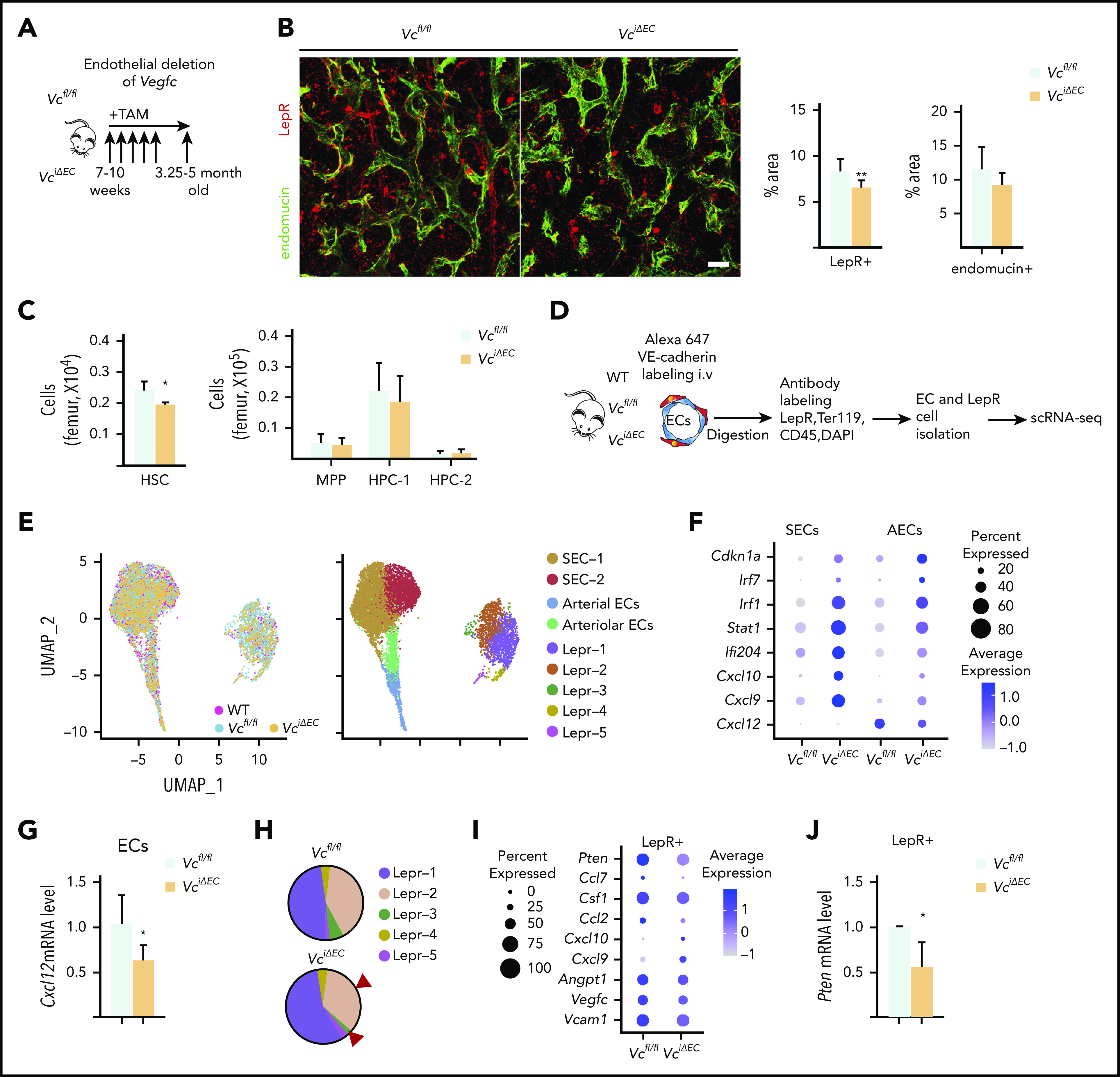
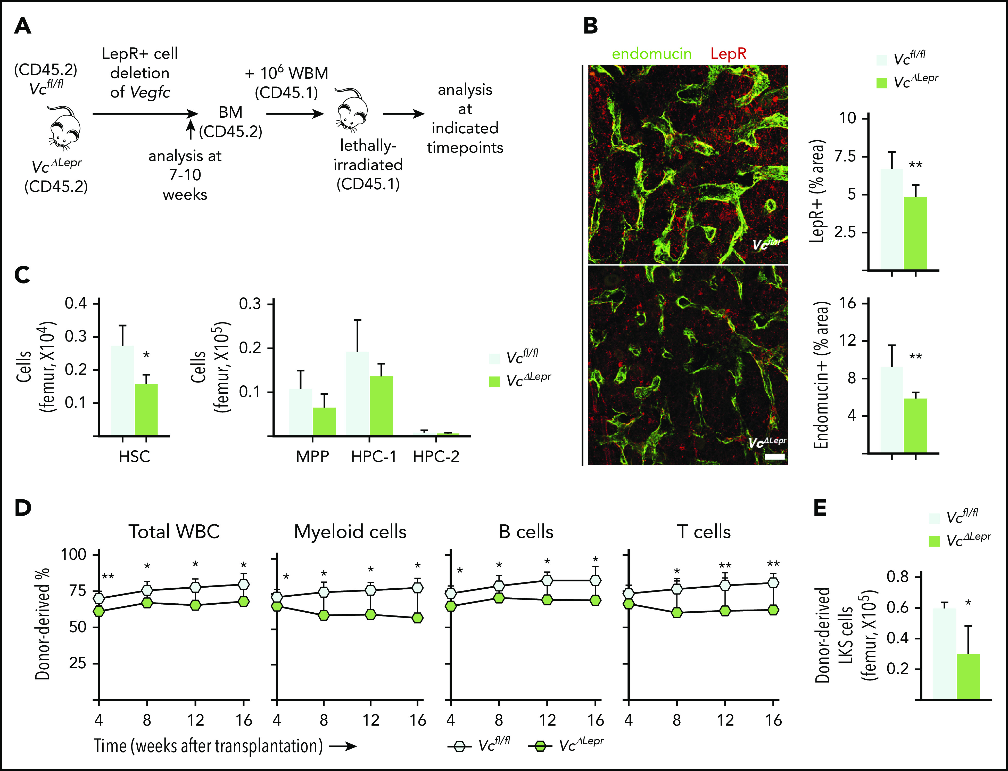
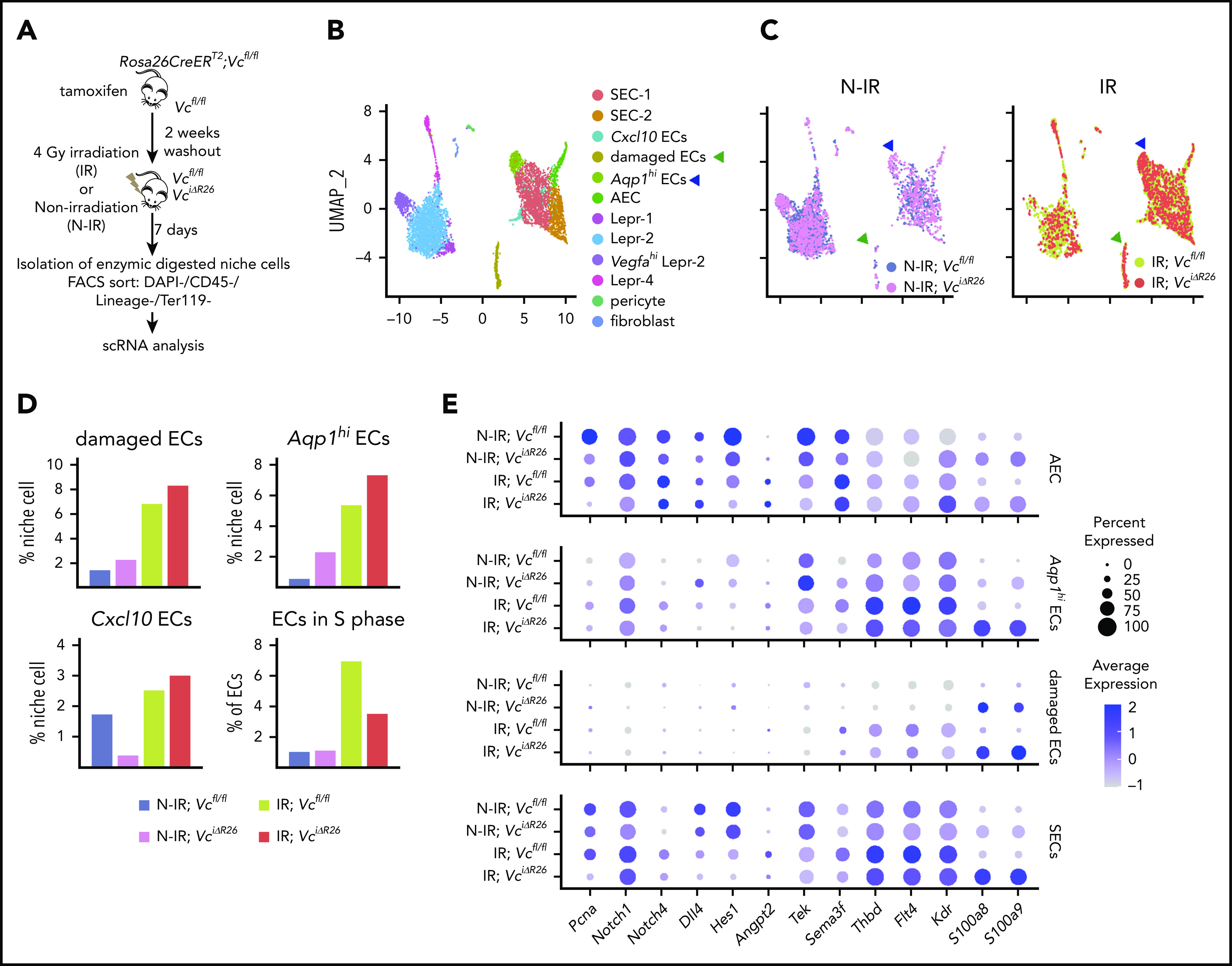
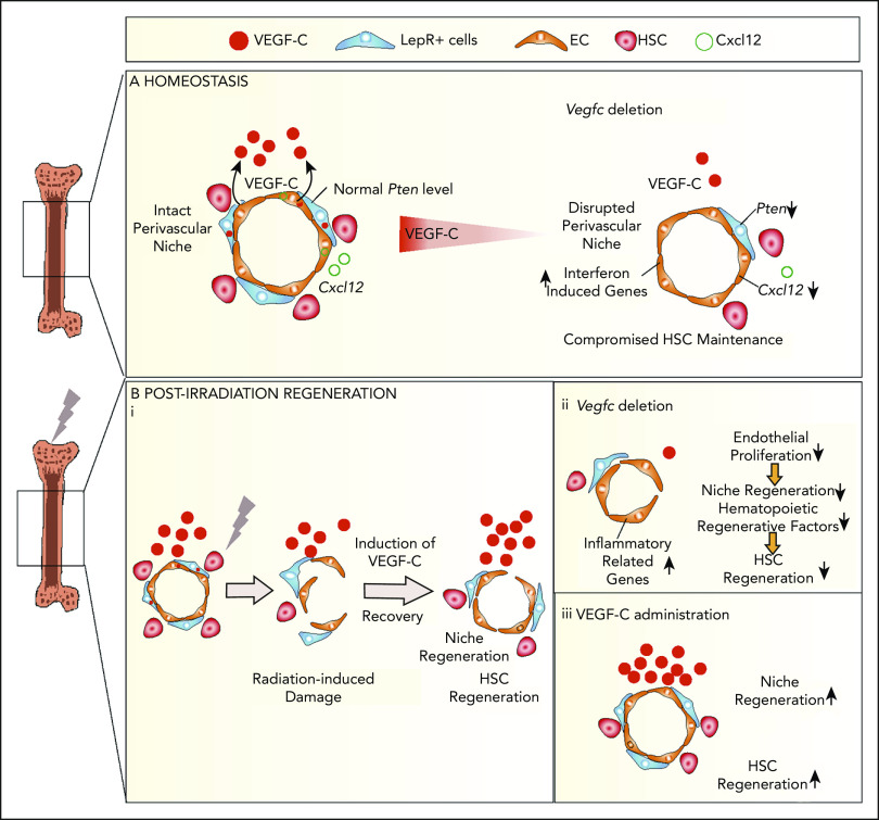
Hematopoietic stem cells (HSCs) reside in the bone marrow (BM) stem cell niche, which provides a vital source of HSC regulatory signals. Radiation and chemotherapy disrupt the HSC niche, including its sinusoidal vessels and perivascular cells, contributing to delayed hematopoietic recovery. Thus, identification of factors that can protect the HSC niche during an injury could offer a significant therapeutic opportunity to improve hematopoietic regeneration. In this study, we identified a critical function for vascular endothelial growth factor-C (VEGF-C), that of maintaining the integrity of the BM perivascular niche and improving BM niche recovery after irradiation-induced injury. Both global and conditional deletion of Vegfc in endothelial or leptin receptor-positive (LepR+) cells led to a disruption of the BM perivascular niche. Furthermore, deletion of Vegfc from the microenvironment delayed hematopoietic recovery after transplantation by decreasing endothelial proliferation and LepR+ cell regeneration. Exogenous administration of VEGF-C via an adenoassociated viral vector improved hematopoietic recovery after irradiation by accelerating endothelial and LepR+ cell regeneration and by increasing the expression of hematopoietic regenerative factors. Our results suggest that preservation of the integrity of the perivascular niche via VEGF-C signaling could be exploited therapeutically to enhance hematopoietic regeneration.


Conflict of interest statement

Conflict-of-interest disclosure: The authors declare no competing financial interests.

Figures

None
Graphical abstract
Figure 1.
Figure 1.
VEGF-C regulates the integrity of the BM LepR+ HSC niche. (A) Relative Vegfc mRNA level in sorted WT PECAM-1+ ECs and LepR+ stromal cells, analyzed by qPCR (normalized to WBM; n = 2 mice per group). (B) Experimental outline of scRNA-seq analysis of LepR+ cells and VE-cadherin–positive ECs from Lepr-Cre;tdTomato BM. (C) UMAP plots of BM LepR-tdTomato–positive cells and VE-cadherin–positive ECs (left). A feature plot shows Lepr and Cdh5 expression (right). (D) A feature plot showing Vegfc, Flt4 (VEGFR-3), and Kdr (VEGFR-2) expression. (E) Experimental setup for evaluating the effects of Vegfc deletion in the BM. Vegfc was deleted from adult BM by administering 5 daily tamoxifen injections to 7- to 10-week-old Rosa26-CreERT2;Vegfcflox/flox mice. (F) Representative confocal immunofluorescence images of femur sections from VciΔR26 and Vcfl/fl mice 5 months after deletion. Staining for ECs (endomucin, green) and perivascular cells (LepR, red), with quantification (right) (n = 5-6 mice per group). Bar represents 50 μm. (G) Quantification of HSPC subsets per femur from 7-month-old VciΔR26 mice and Vcfl/fl littermate controls, determined by CD48 and CD150 staining (n = 13-14 mice per group). Quantification of HSPC subsets per femur from 22- to 23-month-old VciΔR26 mice and Vcfl/fl littermate controls (n = 6 male mice per group). Values show mean ± SD. Statistical significance was determined with the 2-tailed, unpaired Student t test. MPP, multipotential progenitor. *P < .05; **P < .01.
Figure 2.
Figure 2.
Endothelial cells serve as a functionally significant source of VEGF-C in the BM. (A) Experimental setup for evaluating the effects of Vegfc deletion in ECs. Vegfc was deleted from ECs in 7- to 10-week-old Cdh5-CreERT2;Vegfcflox/flox mice by tamoxifen injections. (B) Representative confocal immunofluorescence images of femur sections from VciΔEC mice and their littermate controls stained for ECs (endomucin, green) and LepR+ perivascular cells (red), with quantifications (right) (n = 6 mice per group). Bar represents 50 μm. (C) Quantification of HSPC subsets per femur from Vcfl/fl and VciΔEC mice determined by CD48 and CD150 staining (n = 13-14). (D) Experimental outline of scRNA-seq analysis of LepR+ cells and VE-cadherin–positive ECs from WT, Vcfl/fl, and VciΔEC mice. (E) UMAP plot of integrated BM ECs and LepR+ cells isolated from WT, Vcfl/fl, and VciΔEC mice (left). UMAP plots showing the clustering of integrated BM ECs and LepR+ cells from WT, Vcfl/fl, and VciΔEC mice (right). (F) Dot plot showing selected differentially expression genes in SECs (SEC-1 and -2), AECs, and arteriolar ECs after endothelial Vegfc deletion in comparison with Vcfl/fl mice. (G) Relative Cxcl12 mRNA levels in isolated BM ECs from VciΔEC and littermate control mice analyzed by qPCR. (H) Comparison of relative cell counts in Lepr-1, -2, -3, -4, and -5 clusters from Vcfl/fl and VciΔEC mice. (I) Dot plot showing selected differentially expression genes in LepR+ cells (Lepr-1, -2, and -3) after endothelial Vegfc deletion in comparison with the Vcfl/fl mice. (J) Relative Pten mRNA levels in isolated BM LepR+ cells from VciΔEC and littermate control mice analyzed by qPCR. *P < .05; **P < .01. MPP, multipotential progenitor.
Figure 3.
Figure 3.
LepR+ cell–derived VEGF-C contributes to maintenance of functional HSCs in the BM. (A) Experimental setup for evaluating the effects of LepR+ cell–derived VEGF-C. Vegfc was deleted from LepR+ cells using Lepr-Cre. BM of 7- to 10-week-old mice was analyzed for niche cell and HSC phenotypes. WBM from 10-week-old VcΔLepr or Vcfl/fl mice was transplanted competitively with CD45.1 WBM into lethally irradiated CD45.1 mice. (B) Representative confocal immunofluorescence images of femur sections from VcΔLepr and Vcfl/fl littermate controls stained for ECs (endomucin, green) and LepR+ perivascular cells (red), with quantification (right) (n = 6-7 mice per group). Bar represents 50 μm. (C) Quantification of HSPC subsets per femur from VcΔLepr and Vcfl/fl littermate control mice (n = 4 mice per group) determined by CD48 and CD150 staining. (D) Competitive transplantation of WBM from VcΔLepr and their Vcfl/fl littermate control mice into lethally irradiated WT CD45.1 recipients (2 independent transplants with 3 to 4 recipients per condition per transplant). Shown is a multilineage donor chimerism from peripheral blood at the indicated time points after competitive transplantation. (E) Quantification of LKS cells derived from VcΔLepr or Vcfl/fl mouse BM 16 weeks after transplantation. Values show means ± SD. Statistical significance was determined using the 2-tailed, unpaired Student t test. *P < .05; **P < .01.
Figure 4.
Figure 4.
Loss of VEGF-C from the BM microenvironment delays vascular and HSC regeneration after irradiation. (A) Experimental setup for evaluating Vegfc expression level in the BM after irradiation, by using qPCR. Relative Vegfc mRNA level in WBM, in sorted CD45Ter119 stromal cells, and in CD45+Ter119+ hematopoietic cells 24 hours after 10-Gy radiation (normalized to untreated WBM; n = 2 mice). (B) Experimental setup for evaluating the efficiency of engraftment of WT WBM in lethally irradiated VciΔR26 and Vcfl/fl mice. (C) Transplantation of WT WBM (CD45.1) into lethally irradiated VciΔR26 mice and their Vcfl/fl littermate hosts. Representative flow cytometry graph of total WBCs from peripheral blood 12 weeks after transplantation (left). The kinetics of multilineage donor chimerism from peripheral blood after transplantation (right; n = 6-8 mice per group). (D) Quantification of BM LKS cells in lethally irradiated VciΔR26 mice and their Vcfl/fl littermate recipients 16 weeks after transplantation (n = 6-8 mice per group). (E) Representative confocal immunofluorescence images and quantification. Femur sections from VciΔR26 mice and their littermate controls stained for ECs and basement membranes (endomucin, green; laminin, white) and LepR cells (red) (n = 4-5 mice per group). Bars represent 50 μm. (F) Experimental setup for serial transplantation. WBM (CD45.1) from lethally irradiated primary VciΔR26 and Vcfl/fl recipients was transplanted competitively with CD45.2 WBM into lethally irradiated secondary VciΔR26 and Vcfl/fl recipients. (G) Multilineage donor chimerism from peripheral blood after secondary competitive transplantation (2 independent transplants with 3 recipients per condition per transplant). (H) Quantification of BM LKS numbers in secondary VciΔR26 mice and their Vcfl/fl littermate controls 16 weeks after transplantation. Reported values are mean ± SD. Statistical significance was determined using the 2-tailed, unpaired Student t test. *P < .05; **P < .01.
Figure 5.
Figure 5.
VEGF-C improves BM recovery after irradiation-induced damage. (A) Experimental setup for evaluating the effects of exogenous VEGF-C upon irradiation-induced injury. AAV9 encoding mouse VEGF-C or control protein was injected systemically (IP) to WT mice 7 days before irradiation. BM was analyzed 7 days after irradiation. (B) Quantification of BM VE-cadherin–positive ECs and LepR+ cells in VEGF-C–treated WT mice 7 days after 4-Gy irradiation by flow cytometry (n = 6-8 individual mice per group). (C) Quantification of LKS cells per femur (n = 6-8 mice per group) by flow cytometry. (D) Hematoxylin and eosin staining of femur sections 7 days after 4-Gy irradiation (n = 6-8 mice per group). Bar represents 50 μm. (E) Experimental setup for evaluating the effects of exogenous VEGF-C on the efficiency of hematopoietic engraftment after transplantation. AAV9 encoding mouse VEGF-C or control protein was injected systemically into WT mice 1 day after lethal irradiation and BM transplantation. BM was analyzed 2 and 3 weeks after transplantation. (F-G) Flow cytometry quantification of BM ECs, LepR+ cells, and LKS cells in mice treated with VEGF-C after lethal irradiation and BM transplantation (n = 3 per group per time point). Values show the mean ± SD. Statistical significance was determined using the Student t test or 1-way analysis of variance multiple-comparisons test. *P < .05; **P < .01; ***P < .001.
Figure 6.
Figure 6.
VEGF-C promotes BM EC proliferation and increases the expression of hematopoietic-regenerative factors after irradiation. (A) Experimental setup for evaluating the effects of Vegfc in the BM upon irradiation. Vegfc was deleted from the BM in 10-week-old Rosa26-CreERT2;Vegfcflox/flox mice using tamoxifen injections. VciΔR26 and Vcfl/fl mice received 4-Gy irradiation after a 2-week tamoxifen washout. Niche cells were isolated from irradiated (IR) VciΔR26 and Vcfl/fl mice and their nonirradiated (N-IR) controls 7 days after irradiation and analyzed using scRNA-seq. (B-C) UMAP plot showing the clustering of integrated nonhematopoietic BM stroma cells from IR VciΔR26 and Vcfl/fl mice and their N-IR controls. UMAP plot showing nonhematopoietic BM stroma cells from irradiated VciΔR26 and Vcfl/fl mice and their N-IR controls separately. Note that damaged ECs (green arrowhead) and Aqp1hi ECs (blue arrowhead) are increased after irradiation. (D) Percentages of the clusters (damaged ECs, Aqp1hi ECs, and Cxcl10 ECs) and quantification of ECs in S phase. (E) Dot plot showing selected differentially expressed genes in SECs (SEC-1, SEC-2, and Cxcl10 ECs), damaged ECs, Aqp1hi ECs, and AECs. Analysis of differentially expressed genes was performed between IR;Vcfl/fl vs N-IR;Vcfl/fl and IR;VciΔR26 vs N-IR;VciΔR26.
Figure 7.
Figure 7.
VEGF-C contributes to BM niche maintenance and regeneration after irradiation. Schematic models describing the BM microenvironment of ECs, LepR+ cells, HSCs, and the effects of VEGF-C. (A) During homeostasis, HSCs reside in and are maintained by the intact perivascular niche composed of ECs and LepR+ cells. VEGF-C from both LepR+ cells and ECs maintains an intact BM perivascular niche that is necessary for HSC maintenance. When Vegfc is deleted from ECs or LepR cells, genes related to interferon response are upregulated, and Cxcl12 and Pten are decreased in BM ECs and LepR+ cells, respectively, which leads to niche impairment and compromised HSC maintenance. (Bi) After irradiation and HSC transplantation, Vegfc expression in the niche is upregulated. Microenvironment-derived VEGF-C contributes to endothelial and LepR+ cell regeneration, which in turn is essential for HSC regeneration. (ii) When Vegfc is deleted from the BM microenvironment, genes related to inflammation are upregulated and the proliferation of ECs is decreased, which leads to impaired niche regeneration. Niche-derived hematopoietic regenerative factors are also decreased, resulting in a decreased recovery of HSCs. (iii) Overexpression of VEGF-C improves vascular regeneration, which leads to a better hematopoietic recovery after irradiation or transplantation.

Similar articles

Cited by

References

    1. Morrison SJ, Scadden DT. The bone marrow niche for haematopoietic stem cells. Nature. 2014;505(7483):327-334. - PMC - PubMed
    1. Ramasamy SK, Kusumbe AP, Itkin T, Gur-Cohen S, Lapidot T, Adams RH. Regulation of Hematopoiesis and Osteogenesis by Blood Vessel-Derived Signals. Annu Rev Cell Dev Biol. 2016;32(1):649-675. - PubMed
    1. Gao X, Xu C, Asada N, Frenette PS. The hematopoietic stem cell niche: from embryo to adult. Development. 2018;145(2):dev139691. - PMC - PubMed
    1. Bernad A, Kopf M, Kulbacki R, Weich N, Koehler G, Gutierrez-Ramos JCH. Interleukin-6 is required in vivo for the regulation of stem cells and committed progenitors of the hematopoietic system. Immunity. 1994;1(9):725-731. - PubMed
    1. Sugiyama T, Kohara H, Noda M, Nagasawa T. Maintenance of the hematopoietic stem cell pool by CXCL12-CXCR4 chemokine signaling in bone marrow stromal cell niches. Immunity. 2006;25(6):977-988. - PubMed

MeSH terms