. 2020 Aug 23;21(17):6066.
doi: 10.3390/ijms21176066.

In Vitro Maturation of Retinal Pigment Epithelium Is Essential for Maintaining High Expression of Key Functional Genes

Affiliations

In Vitro Maturation of Retinal Pigment Epithelium Is Essential for Maintaining High Expression of Key Functional Genes

Abdullah Al-Ani et al. Int J Mol Sci. .

Abstract

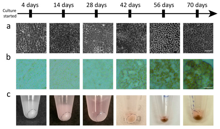
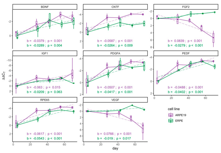
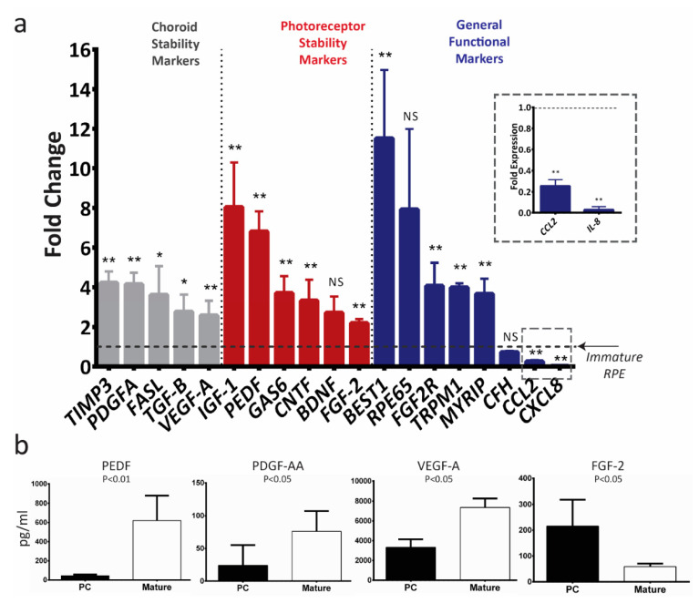
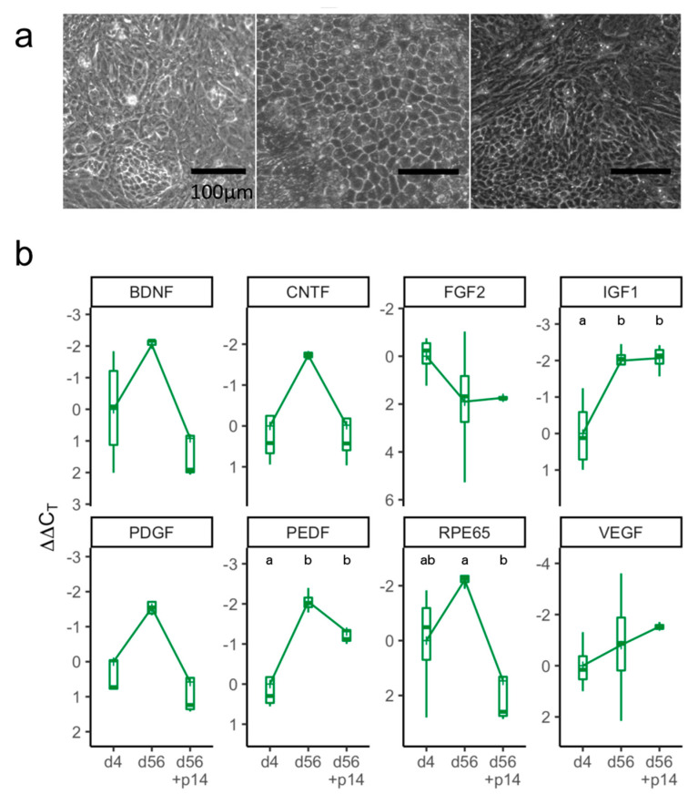
Age-related macular degeneration (AMD) is the leading cause of blindness in the industrialized world. AMD is associated with dysfunction and atrophy of the retinal pigment epithelium (RPE), which provides critical support for photoreceptor survival and function. RPE transplantation is a promising avenue towards a potentially curative treatment for early stage AMD patients, with encouraging reports from animal trials supporting recent progression toward clinical treatments. Mature RPE cells have been reported to be superior, but a detailed investigation of the specific changes in the expression pattern of key RPE genes during maturation is lacking. To understand the effect of maturity on RPE, we investigated transcript levels of 19 key RPE genes using ARPE-19 cell line and human embryonic stem cell-derived RPE cultures. Mature RPE cultures upregulated PEDF, IGF-1, CNTF and BDNF-genes that code for trophic factors known to enhance the survival and function of photoreceptors. Moreover, the mRNA levels of these genes are maximized after 42 days of maturation in culture and lost upon dissociation to single cells. Our findings will help to inform future animal and human RPE transplantation efforts.

Keywords: cell culture; differentiation; embryonic stem cells; maturation; pigment epithelium derived factor (PEDF); retinal pigment epithelium (RPE).


Conflict of interest statement

The authors declare no conflict of interest.

Figures

Figure 1
Figure 1
Embryonic stem cell-derived retinal pigment epithelium (E-RPE) cultures demonstrate morphological changes and become pigmented as they mature. (a) Cellular morphology of E-RPE cultures develops over time to make the consistent hexagonal shaped cells seen at 56 and 70 days. (b) Representative micrographs of the progressive pigmentation of the E-RPE culture as it matures. (c) Cell pellets in microcentrifuge tubes of E-RPE cultures sampled at confluency (4 days), 14 days, 28 days, 42 days, 56 days and 70 days show increasing pigmentation over time. Scale bars represent lengths of 50 µm in (a) and (b).
Figure 2
Figure 2
Key functional genes progressively increase with maturation. Real-time polymerase chain reaction (RT-qPCR) analysis of mRNA levels across 70 days of culture in E-RPE culture (green) and ARPE-19 culture (purple). Results were normalized to an endogenous reference gene (PPIA) and are presented as ΔΔCt means (n = 4) ± standard deviation at each time point. A linear regression model (dashed line) was used to describe the relationship between ΔΔCt values and days of maturation of each gene for both RPE cell sources; regression coefficients, denoted as b, and associated p-values, to reject the null hypothesis that b = 0, are shown on each graph.
Figure 3
Figure 3
In vitro maturation of ARPE-19 upregulates the expression of therapeutically relevant photoreceptor and choroid trophic factors. (a) RT-qPCR analysis of mRNA levels of key RPE genes in mature ARPE-19 cells (>42 days in culture; n = 4). The results were normalized to an endogenous reference gene (PPIA) and are presented as mean fold change (2ΔΔCT) relative to immature ARPE-19 cultures (dotted line) ± standard deviation. Data were compared with a Mann–Whitney U test of ΔCt values; * p < 0.05; NS: not significant. (b) Protein secreted by the post-confluent (PC) immature and mature ARPE-19 cultures into the conditioned media presented as mean concentration ± standard deviation (n = 5), Mann–Whitney U test.
Figure 4
Figure 4
In vitro maturation of E-RPE cells upregulates the expression of therapeutically relevant photoreceptor and choroid trophic factors. (a) RT-qPCR analysis of mRNA levels of key RPE genes in mature E-RPE cells (>42 days in culture; n = 5). The results were normalized to an endogenous reference gene (PPIA) and are presented as mean fold change (2ΔΔCT) relative to immature E-RPE culture (dotted line) ± standard deviation. Data were compared with a Mann–Whitney U test of ΔCt values; * p < 0.05; ** p < 0.01; NS: not significant. (b) Protein secreted by the post-confluent (PC) immature and mature E-RPE cultures into the conditioned media presented as mean concentration ± standard deviation (n = 5), Mann–Whitney U test.
Figure 5
Figure 5
Markers of maturation are lost after passaging E-RPE cells. (a) Morphology of E-RPE cells at confluence, four days after seeding (left), at 56 days of maturation (center) and at a further 14 days after passaging (right). Scale bar represents 100 μm. (b) RT-qPCR analysis at three time points (4 days, 56 days and 56 + 14 days after passaging). Results were normalized to PPIA and represent mean ΔΔCt values (n = 3 or 4) ± standard deviation plotted on a negative y-axis (higher expression at the top). Kruskal-Wallis one-way ANOVA test was used to compare ΔΔCt values of the various maturation points within each gene followed by a Tukey’s honest significance test; different letters, p < 0.05.

References

    1. Wong W.L., Su X., Li X., Cheung C.M.G., Klein R., Cheng C.-Y., Wong T.Y. Global prevalence of age-related macular degeneration and disease burden projection for 2020 and 2040: A systematic review and meta-analysis. Lancet Glob. Heal. 2014;2:e106–e116. doi: 10.1016/S2214-109X(13)70145-1. - DOI - PubMed
    1. Chou C.-F., Cotch M.F., Vitale S., Zhang X., Klein R., Friedman D.S., Klein B.E., Saaddine J.B. Age-Related eye diseases and visual impairment among U.S. adults. Am. J. Prev. Med. 2013;45:29–35. doi: 10.1016/j.amepre.2013.02.018. - DOI - PMC - PubMed
    1. Frick K.D., Gower E.W., Kempen J.H., Wolff J.L. Economic Impact of Visual Impairment and Blindness in the United States. Arch. Ophthalmol. 2007;125:544. doi: 10.1001/archopht.125.4.544. - DOI - PubMed
    1. Rein D.B., Zhang P., Wirth K.E., Lee P.P., Hoerger T.J., McCall N., Klein R., Tielsch J.M., Vijan S., Saaddine J. The Economic Burden of Major Adult Visual Disorders in the United States. Arch. Ophthalmol. 2006;124:1754–1760. doi: 10.1001/archopht.124.12.1754. - DOI - PubMed
    1. Lim L.S., Mitchell P., Seddon J.M., Holz F.G., Wong T.Y. Age-Related macular degeneration. Lancet. 2012;379:1728–1738. doi: 10.1016/S0140-6736(12)60282-7. - DOI - PubMed

LinkOut - more resources